Technology Platforms
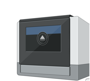
10x Genomics Chromium iX Controller

Droplet-based (10x Genomics) single-cell RNA sequencing enables high-throughput analysis of individual cells. Comprehensive information on the global gene expression profiles of large numbers of individual cells—derived, for example, from digested tissues or blood—is generated. Single cells are encapsulated together with reagents into tiny aqueous droplets in an oil flow using a microfluidic system. Within each droplet, mRNA is isolated, reverse transcribed into cDNA, and labeled with a unique barcode that identifies each cell. All cDNAs are then sequenced in a single sequencing run.
This technology enables the resolution of heterogeneity within seemingly homogeneous cell populations, the classification and characterization of previously unknown cell types, and provides insights into biological development and differentiation processes at the single-cell level. It is particularly valuable for investigating inflammatory processes and immune responses, including tumor-infiltrating immune cells.
Flow cytometry

•BD FACSymphony™ A1 Cell Analyzer
•BD FACSymphony™ A3 Cell Analyzer
•BD LSRFortessa™ Cell Analyzer
•BD FACSAria™ III Cell Sorter
Flow cytometry and FACS (fluorescence activated cell sorting) are fast and reliable methods for characterizing and isolating specific cell subpopulations based on fluorescent labeling. Our platform supports a wide range of applications across different organisms and cell types.
We offer state-of-the-art infrastructure and expert support for a broad spectrum of cell analysis and sorting applications. The high-performance cytometers enable simultaneous measurement of a large number of parameters, providing detailed insights into cellular functions and states. Moreover, the Aria III system allows for precise sorting of cell populations and single-cell deposition for downstream analyses.
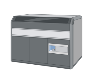
Imaging Flow Cytometry
This technology combines the quantitative power of flow cytometry with high-resolution imaging, enabling the analysis of cell populations while simultaneously capturing detailed fluorescence and morphological data at the single-cell level. It is particularly well-suited for studying complex cellular processes such as signal transduction, protein localization, cell-cell interactions, and apoptosis.
With the ability to capture both brightfield and fluorescence images, the system allows rapid analysis of thousands of cells, providing statistically robust data with single-cell resolution. To effectively analyze the complex data generated by imaging flow cytometry (IFC), we have developed MorphoMapping—an analytical framework that utilizes advanced techniques such as dimensionality reduction and clustering. This tool enables automated identification of patterns, causal relationships, and previously unrecognized cell populations.
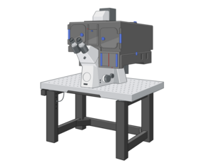
Confocal and Superresolution - Fluorscene Microscopy (TIRF)

Our state-of-the-art fluorescence microscopy system combines confocal microscopy, structured illumination microscopy (SIM), and total internal reflection fluorescence microscopy (TIRF) in a single device. This versatile platform enables precise visualization of cellular and subcellular structures, as well as dynamic processes, with extremely high resolution and specificity.
Confocal microscopy provides high-contrast, three-dimensional images of cells and tissues. High resolution is achieved by detecting light only from the focal plane, while out-of-focus light is excluded. This makes it particularly suitable for analyzing complex specimens such as tissue sections or organoids. SIM overcomes the diffraction limit of light and achieves nanometer-scale precision (super-resolution), revealing the finest cellular details. TIRF, on the other hand, allows highly sensitive investigation of processes occurring in the immediate vicinity of the plasma membrane by exciting fluorescence only within a thin region (~100–200 nm) above the membrane. This technique is ideal for studying membrane-associated processes, molecular interactions, and vesicle trafficking.
Using this combined system, we analyze dynamic processes such as the organization of immunological synapses, redistribution of signaling proteins during leukocyte activation, vesicular transport, and the actin cytoskeleton. By integrating confocal, super-resolution, and TIRF microscopy into a single platform, we can not only investigate structural details at the subcellular level but also track molecular processes in real time.
Combining these microscopy techniques with other analytical approaches such as imaging flow cytometry opens new avenues for exploring immune cell behavior across multiple levels—from population analysis down to subcellular resolution. This platform is of exceptional value for elucidating the regulation of the immune system and cellular processes in health and disease.
