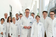

Musculoskeletal Imaging Team
The section for MSK Radiology aims to provide high-end imaging and image-guided intervention of the entire musculoskeletal system including the spine. Using the latest generation of a 3 Tesla MR scanner a special research focus is to evaluate new high resolution and highly accelerated MR sequences that offer improved image quality at decreased scanning time. These include AI-powered image reconstruction technologies with a deep neural network at its core. Another focus is the field of compositional or biochemical MR imaging. With the use of special MR techniques like T2 mapping or dGEMRIC it is possible to detect and quantify the ultrastructure and the biochemical composition of tissues. We have successfully used these techniques in previous projects to evaluate the cartilage properties of several joints and have detected early ultrastructural damage (early osteoarthritis) and changes (healing) after therapy. Current and future efforts will concentrate on biochemical analyses of other tissues, for instance the glenoid labrum and the biceps tendon and others. Also, possible future developments regarding the biochemical sequence techniques will be established and evaluated.
Another focus of the section is high-precise and low-dose CT guided intervention. Since years we provide and develop advanced CT guided interventions for pain therapy, bone and soft tissues biopsies up to radiofrequency ablation (RFA) of osteoid osteoma and even osteoblastoma. The 2022 installed “top of the line” single-source CT scanner (Siemens Somatom X.ceed) offers a plenty of new techniques and developments to further improve these interventions. These include techniques to significantly reduce radiation dose and also to provide ultra-high-resolution imaging including improved target and needle imaging during the intervention. The newly available gantry-integrated laser guidance and the MR-CT image fusion aims to further improve high precision high yield biopsy and therapy and will be evaluated in future projects.
Tissue composition analyses using MRI T2 mapping
Patrick Stein, Christoph Rehnitz
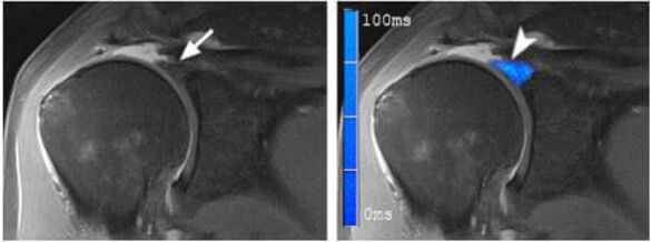
MRI T2 mapping can detect and quantify early degenerative changes in articular cartilage, such as proteoglycan loss, collagen disruption, and increased water content. In the current project, we are investigating the possibility to assess the integrity of other structures than the cartilage like the glenoid labrum (see Fig. 1) and the long biceps tendon. In an arthroscopy controlled study at the shoulder we aim to characterize normal tendons and labra as well as different degrees of pathology. Future efforts will focus on translating these results to other joints and structures and improvements/acceleration of the sequence.
Improvement of cartilage imaging in patients after cartilage repair using deep learning-based image quality improvements and acquisition acceleration.
Patrick Stein, Christoph Rehnitz
Deep learning (DL) based algorithms are trained on large datasets of MRI images and can automatically extract complex features and patterns from the data. This led to newly available DL and AI based sequences which potentially lead to improved image quality, tissue sharpness, resolution and acquisition speed. We aim to evaluate these techniques in different patient settings including cartilage repair.
Establishment of a low-dose protocol for CT-guided periradicular therapy (PRT) in patients suffering from lumbar pain
Thomas Schneider, Christoph Rehnitz
Objectives: Reducing radiation exposure while maintaining diagnostic and therapeutic efficacy of PRT.
Materials and methods: Implementation of state-of-the-art technology such as the high end detector (128 acquired slices, Z-Sharp-technique, decreased cross-talk and noise levels), tin filter technique, modern iterative reconstruction algorithms. Active, protocol-guided adjustments of scan parameters by radiologist/ radiological technologist.
Establishment of a low-dose protocol with highly precise needle guidance for CT-guided radiofrequency ablation of osteoid osteomas
Thomas Schneider, Christoph Rehnitz
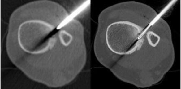
Objectives: To reduce radiation exposure in CT-guided radiofrequency ablation of osteoid osteomas while optimizing the precision of ablation positioning with consistent or improved therapeutic outcome in patients suffering from osteoid osteoma.
Materials and methods:
UHR-comb is employed for focused utilization of ultra-high resolution (UHR) imaging to reduce hardening artifacts associated with the ablation materials, resulting in optimized, central positioning within the nidus while maintaining radiation dose level lower than conventional intervention mode.
Implementation of state-of-the-art technology such as the high end detector (128 acquired slices, Z-Sharp-technique, decreased cross-talk and noise levels), tin filter technique, modern iterative reconstruction algorithms.
Active, protocol-guided adjustments of scan parameters by radiologist/ radiological technologist.
High precision laser guided biopsy of bone tumors with MR/CT fusion
Maike Küchler, Thomas Schneider, Christoph Rehnitz
The availability of a gantry-integrated laser guidance offers the potential of a more precise needle guidance in reduced intervention time. The fusion of MR and CT images during the intervention enables to adjust the biopsy path based on MR findings (e.g. diffusion restricted areas) that are not visible with CT imaging alone, possibly leading to improved biopsy results. Current status: clinical establishment and evaluation of true potential in clinical practice.
Selected publications
1. Stein P, Wuennemann F, Schneider T, Zeifang F, Burkholder I, Weber MA, Kauczor HU, Rehnitz C. 3-Tesla T2 Mapping Magnetic Resonance Imaging for Evaluation of SLAP Lesions in Patients with Shoulder Pain: An Arthroscopy-Controlled Study. J Clin Med. 2023 Apr 25;12(9):3109.
2. Heiss R, Weber MA, Balbach E, Schmitt R, Rehnitz C, Laqmani A, Sternberg A, Ellermann JJ, Nagel AM, Ladd ME, Englbrecht M, Arkudas A, Horch R, Guermazi A, Uder M, Roemer FW. Clinical Application of Ultrahigh-Field-Strength Wrist MRI: A Multireader 3-T and 7-T Comparison Study.Radiology. 2023 Apr;307(2):e220753
3. Kintzelé L, Brandelik SC, Wuennemann F, Weber MA, Lehner B, Kauczor HU, Rehnitz C. MRI patterns indicate treatment success and tumor relapse following radiofrequency ablation of osteoblastoma. Int J Hyperthermia. 2020;37(1):274-282.
4. Rehnitz C, Kuni B, Wuennemann F, Chloridis D, Kirwadi A, Burkholder I, Kauczor HU, Weber MA. Delayed gadolinium-enhanced MRI of cartilage (dGEMRIC) and T2 mapping of talar osteochondral lesions: Indicators of clinical outcomes. J Magn Reson Imaging. 2017 Dec;46(6):1601-1610.
5. Rehnitz C, Kupfer J, Streich NA, Burkholder I, Schmitt B, Lauer L, Kauczor HU, Weber MA. Comparison of biochemical cartilage imaging techniques at 3 T MRI. Osteoarthritis Cartilage. 2014 Oct;22(10):1732-42.