
Welcome
Dear Patients and Visitors,
Welcome to the Center for Skull Base Diseases at Heidelberg University Hospital. Our center is dedicated to the expert care of individuals with conditions affecting the skull base, including tumors and related disorders. With our commitment to state-of-the-art treatment methods, decades of experience, and care tailored to each individual, our team is here to support you at every stage.
Whether you require tumor treatment, reconstructive surgery, or access to innovative therapies – we are committed to making your stay as effective and comfortable as possible. Our specialists work closely with experts from other disciplines to provide you with comprehensive, personalized care that is aligned with your individual needs and treatment goals.
Sincerely,
Prof. Dr. Dr. Dr. h.c. Jürgen Hoffmann, Director of the Department of Oral and Maxillofacial Surgery
Univ.-Prof. Dr. med. Sandro Krieg, Director of the Department of Neurosurgery
Prof. Dr. med. Patrick Schuler, Director of the Department of Otorhinolaryngology
Prof. Dr. med. Dr. rer. nat. Jürgen Debus, Director of the Department of Radiation Oncology
Involved Specialties
Oral and Maxillofacial Surgery
Oral and Maxillofacial Surgery specializes in the surgical treatment of diseases, injuries, and malformations of the mouth, jaws, face, and skull base. This includes tumor removal, complex reconstructions, as well as aesthetic and functional restorations. Using advanced surgical expertise with state-of-the-art technology to ensure safe, precise, minimally-invasive and patient-centered care.
Neurosurgery
Neurosurgery is dedicated to the surgical treatment of disorders of the central and peripheral nervous system. This includes procedures involving the brain, spinal cord, and nerves, with a particular focus on the complex anatomy of the skull base. Technological innovations such as navigation systems, intraoperative imaging, and minimally invasive techniques are integral parts of modern neurosurgery.
Radiation Oncology
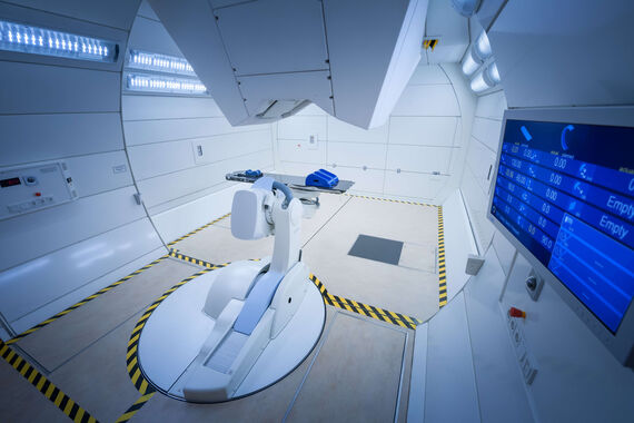
Radiation oncology focuses on the treatment of cancer through the targeted use of ionizing radiation. The goal is to precisely destroy or control tumor tissue while sparing healthy tissue as much as possible. Cutting-edge methods such as proton therapy and image-guided radiation techniques are employed. As one of only a few centers, irradiation with heavy ions is also available at HIT (Heidelberg Ion-Beam Therapy Center).
Otorhinolaryngology (ENT)
Otorhinolaryngology covers the diagnosis and treatment of diseases in the head and neck region, including the skull base. This includes both inflammatory conditions as well as benign and malignant tumors. In addition to surgical procedures, the ENT department also offers reconstructive and function-preserving treatment approaches.
About the Skull Base Center
Comprehensive Expertise
Our clinic specializes in the treatment of a wide range of conditions, including injuries, congenital malformations, head and neck tumors, as well as complex reconstructive procedures. The team is led by internationally recognized surgeons with extensive experience in managing both routine cases and particularly challenging situations.
Interdisciplinary Approach
Our clinic works closely with specialists from other departments of Heidelberg University Hospital to ensure comprehensive care that takes all aspects of each patient’s condition into account. This integrated approach enables us to develop optimal, individualized treatment plans, particularly for complex conditions.
State-of-the-Art Technology
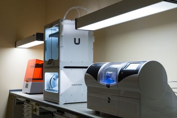
Heidelberg University Hospital is equipped with the latest surgical technologies, including 3D imaging and computer-assisted surgery. These innovations enable highly precise and minimally invasive procedures. The use of such technologies contributes to improved treatment outcomes, shorter recovery times, and optimized aesthetic results. In addition, cutting-edge radiation therapies—such as heavy ion irradiation at the Heidelberg Ion-Beam Therapy Center (HIT)—are available.
Research and Innovation
As part of Heidelberg University Hospital, one of Europe’s leading medical research institutions, our clinic is actively involved in clinical research and trials. This allows our patients to benefit from the latest advances in oral and maxillofacial surgery. At the same time, we contribute significantly to the further development of medical knowledge and modern treatment methods.
Which conditions are treated at the Skull Base Center?
At the Skull Base Center of Heidelberg University Hospital, both benign and malignant conditions of the skull base are treated through an interdisciplinary approach. These include tumors and other disorders that originate directly at the skull base or affect its structures. Typical conditions we treat include:
- Tumors of the pituitary gland (hypophysis), such as pituitary adenomas and craniopharyngiomas
- Benign tumors of the meninges, so-called meningiomas, occurring at the anterior, middle, or posterior skull base
- Benign tumors of the vestibular nerve, known as vestibular schwannomas (also called acoustic neuromas)
- Rare malignant tumors, such as chordomas or sarcomatous tumors of the skull base
- Tumors of the nasal cavity and paranasal sinuses that may infiltrate the skull base, including squamous cell carcinoma, adenoid cystic carcinoma, and olfactory neuroblastoma
- Diseases of the temporal bone, e.g. cholesteatomas extending toward the skull base
- Tumors of the eye socket (orbit)
- Rare skull base tumors, such as glomus tumors and vascular anomalies
- Inflammatory processes, such as osteomyelitis or osteoradionecrosis of the skull base
- Skull base injuries, e.g. after accidents or as a result of previous treatments
- Malformations of the skull base as well as spontaneous or post-therapeutic cerebrospinal fluid leaks (rhinorrhea or otorrhea)
Thanks to the close collaboration between the departments of Neurosurgery, Otorhinolaryngology (ENT), Oral and Maxillofacial Surgery, and Radiation Oncology, we provide comprehensive, specialized diagnostics and therapy from a single source. Depending on the condition, we develop an individualized treatment plan – ranging from surgical procedures and radiation or chemotherapy to combined, innovative therapeutic approaches.
What Are Skull Base Tumors?
Skull base tumors are a diverse group of neoplasms that develop in the complex region at the base of the skull—where the brain rests and numerous important nerves and blood vessels pass through. These tumors can be benign or malignant and may originate from different tissues such as bone, nerves, or glands. Because of their proximity to vital structures, even benign tumors can cause significant symptoms and therefore require specialized treatment.
Common Types of Skull Base Tumors
Meningiomas:
The most common benign tumors of the skull base, meningiomas arise from the meninges, the membranes covering the brain and spinal cord. Although they are benign, they may grow large enough to compress surrounding structures and cause various neurological symptoms.
Pituitary Adenomas:
These tumors develop in the pituitary gland, located at the skull base. They are usually benign but can cause hormonal disorders, visual disturbances, or pressure symptoms due to their location close to the optic nerves and other critical structures.
Vestibular Schwannomas (Acoustic Neuromas):
Benign tumors that arise from Schwann cells surrounding the vestibular nerve, which connects the inner ear to the brain. Typical symptoms include hearing loss, tinnitus (ringing in the ear), and balance problems.
Chordomas:
Rare malignant tumors that originate from remnants of the notochord, a structure important in early spinal development. Chordomas grow slowly but can destroy surrounding bone and soft tissue.
Sinonasal Carcinomas:
Malignant tumors that arise in the nasal cavity or paranasal sinuses and may extend into the skull base. Typical symptoms include nasal obstruction, facial pain, and swelling.
Symptoms
The symptoms of skull base tumors vary depending on their size, location, and type. Common symptoms include:
- Severe, persistent headaches
- Visual disturbances (double vision, vision loss)
- Hearing loss or tinnitus
- Balance or coordination problems
- Numbness or weakness in the face
- Hormonal disorders (e.g., with pituitary adenomas)
Because these tumors can compress critical structures such as cranial nerves or the brainstem, early diagnosis is essential to prevent permanent neurological damage.
Treatment Options
Treatment depends on the type, location, and size of the tumor as well as the patient’s overall health. Common approaches include:
Surgical Removal:
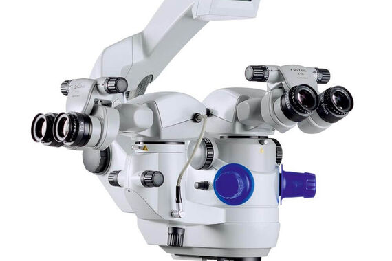
For most skull base tumors, surgery is the primary treatment option. Minimally invasive techniques, such as endoscopic or microscopic surgery, allow access to the tumor while minimizing damage to surrounding tissue. The use of modern tools such as surgical navigation and intraoperative imaging increases the safety and precision of these procedures.
Radiation Therapy:
Often used for inoperable tumors or as an adjunct to surgery. Techniques such as stereotactic radiosurgery (e.g., Gamma Knife) allow for high-dose irradiation of the tumor while sparing healthy tissue.
Chemotherapy:
Less commonly used, but may be appropriate for certain malignant skull base tumors, particularly aggressive or metastatic types.
Observation:
In some cases—especially with small, benign tumors that cause no symptoms—close monitoring with regular imaging may be the best approach.
Conclusion
Skull base tumors represent a complex medical challenge due to their location and potential impact on vital structures. Early diagnosis and an interdisciplinary approach involving neurosurgeons, ENT specialists, and oncologists are crucial to achieving the best possible treatment and prognosis.
What Is Modern Skull Base Surgery?
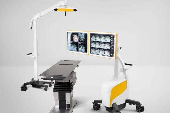
Comprehensive and interdisciplinary skull base surgery is a highly specialized field within neurosurgery and otorhinolaryngology (ENT), requiring close collaboration across multiple medical disciplines. The complex anatomy of the skull base, which contains numerous vital neurovascular structures, demands a deep understanding of anatomy, precise surgical planning, and the use of modern technologies to ensure maximum safety and effectiveness.
In recent years, technological advances have significantly transformed skull base surgery, particularly through the integration of navigation systems and intraoperative imaging. These innovations have improved the accuracy of tumor removal, reduced complications, and greatly enhanced treatment outcomes for patients.
Navigation systems, often compared to a GPS for the human body, have become indispensable in skull base surgery. They allow surgeons to map a patient’s anatomy in detail prior to surgery using high-resolution imaging techniques such as CT and MRI. During the procedure, the navigation system tracks the position of surgical instruments in real time and continuously matches them with preoperative imaging data. This is especially crucial in skull base surgery, where millimeter-level precision is required to avoid injury to surrounding structures such as cranial nerves, blood vessels, or the brainstem.
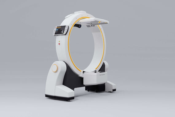
The integration of intraoperative imaging further enhances surgical precision. Techniques such as intraoperative CT or MRI provide real-time imaging during surgery. This is particularly valuable for assessing the extent of tumor resection and immediately identifying and removing any remaining tumor tissue. By providing up-to-date images during the procedure, the risk of leaving behind residual tumor tissue that is difficult to detect with conventional visual inspection is minimized. In addition, intraoperative imaging facilitates navigation in cases of complex anatomical variations and helps verify critical surgical steps, such as the integrity of the dura mater or the completeness of skull base reconstruction.
Another modern technique is the use of 3D modeling and 3D printing. Patient-specific models of the skull base can be created before surgery, supporting precise surgical planning and enabling surgeons to simulate complex procedures in advance, better anticipating potential challenges.
The success of skull base surgery relies heavily on interdisciplinary collaboration. Neurosurgeons, ENT specialists, radiologists, and in some cases plastic surgeons work hand in hand, combining their expertise—particularly in the treatment of complex tumors, vascular malformations, or trauma affecting the skull base.
Conclusion
Modern technologies, especially navigation systems and intraoperative imaging, have revolutionized skull base surgery. Today, they enable more precise, safer, and more effective procedures. The future of skull base surgery lies in continued technological innovation and in the advancement of minimally invasive and highly precise surgical techniques.
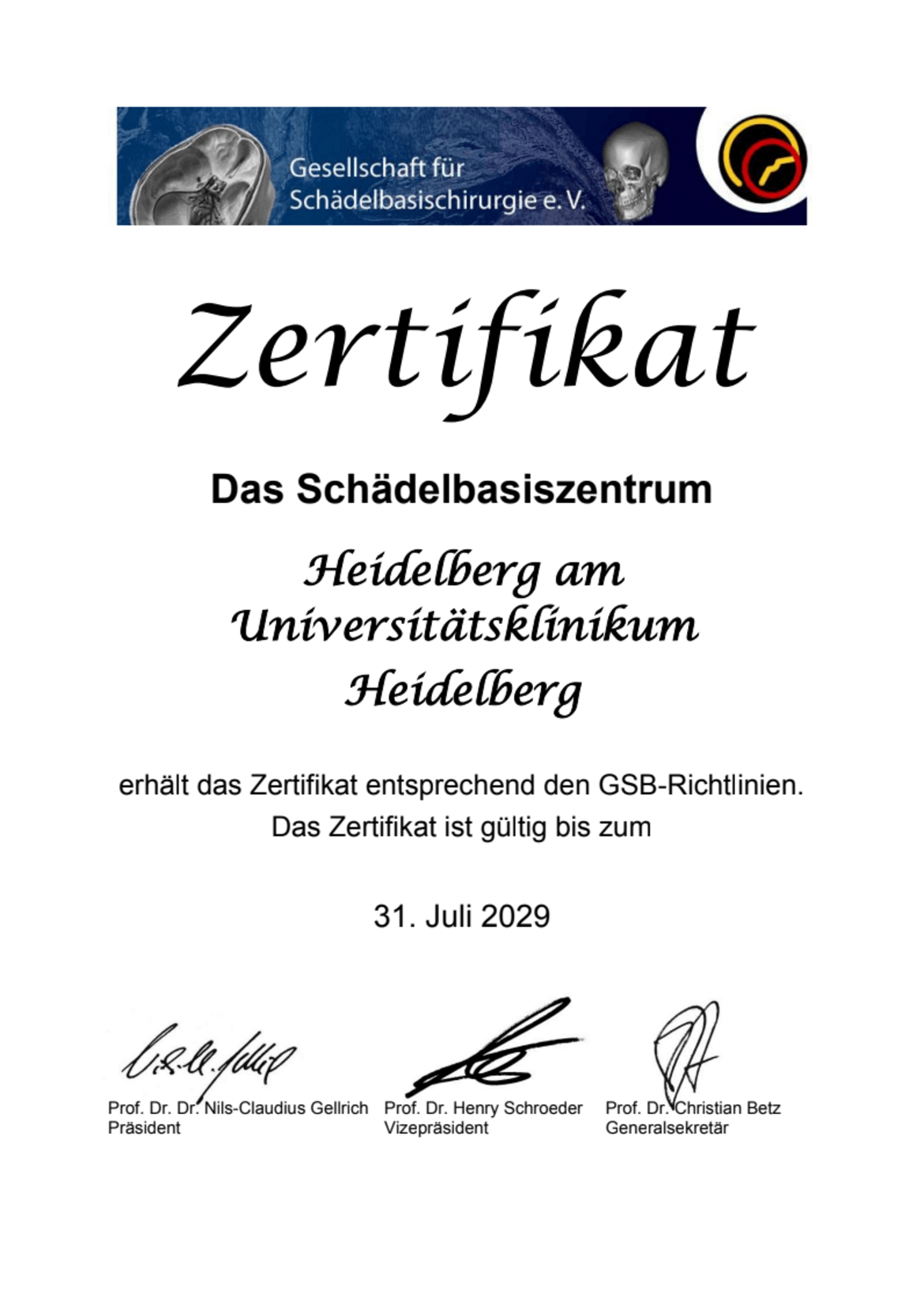
Certified Skull Base Center
The Skull Base Center at Heidelberg University Hospital has been officially certified by the German Society for Skull Base Surgery (GSB). This certification confirms that our center meets the high quality standards and comprehensive requirements set forth in the GSB guidelines. It particularly recognizes the interdisciplinary collaboration of our departments as well as our outstanding expertise in the diagnosis and treatment of complex skull base diseases.
The certificate has been awarded until July 31, 2029, underscoring our commitment to providing patients with specialized, high-quality care based on the latest scientific knowledge. The certification is founded on structured, coordinated, and patient-centered care that combines state-of-the-art medical technology with decades of clinical experience.
We are proud of this recognition and see it as motivation to continuously advance our dedication to the treatment of skull base diseases.
Patient Registration at the Skull Base Center
If you have been diagnosed with a condition affecting the skull base, or if there is a corresponding suspicion, you can directly consult the department responsible for your condition (Neurosurgery, Oral and Maxillofacial Surgery, Otorhinolaryngology, or Radiation Oncology).
Please bring along any existing medical records, imaging (CT/MRI), and physician reports to your appointment so that we can comprehensively assess your condition.
Service for International Patients
At Heidelberg University Hospital, we understand that receiving medical treatment abroad can be a major challenge. Our International Office supports international patients at every stage of their stay and offers the following services:
- Individual treatment plans: Our surgeons develop a therapy plan tailored to your specific condition and personal needs.
- Multilingual support: We provide language assistance and interpreting services to ensure clear and smooth communication throughout the entire treatment process.
- Logistical assistance: The International Office helps you and your family with travel arrangements, accommodation, and other supportive services.
Contact
If you are considering treatment at the Skull Base Center of Heidelberg University Hospital, our team will be happy to assist you. Please contact the International Office for further information or to arrange a consultation with one of our specialists.