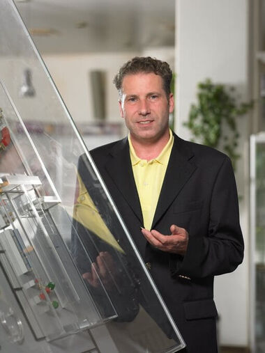
Interview with Professor Thomas Haberer, PhD

Science-Technical Director of HIT
Interview from November 2009
At the Heidelberg Ion-Beam Therapy Center HIT, ion beams are used to treat cancer patients. What are ions? What ions are used at HIT?
Ions are atoms from whose shell electrons have been removed. These ions are thus positively charged and can be accelerated and guided in electromagnetic fields. At HIT protons (ions of hydrogen atoms) and helium, carbon, and oxygen ions are used. In comparison with the photons used in conventional radiotherapy, protons and helium ions allow much more precise irradiation of the area being treated and thus much less radiation damage to healthy tissue, in particular organs at risk. In addition to the increased precision in localizing the distribution of the dose, carbon and oxygen ions have a much greater efficacy, especially at the end of their range, i.e. in the tumour tissue. For the first phase of patient treatment, protons and carbon ions are used.
How are the ions generated at HIT?
The HIT accelerator system has two ion sources that currently generate protons and carbon ions in parallel operation. The central element of the ion source is the plasma chamber, where magnetic fields trap the ions and electrons in circular paths and microwaves are overlaid for more efficient heating. An initially neutral gas is introduced into the plasma chamber. Due to heating, the electrons that are released from the atomic shell are accelerated and consecutively ionize other gas molecules. The positively charged ions are then extracted from the plasma that arises in the chamber by applying high voltage.
What happens in the linear accelerator?
In the linear accelerator, particle bundles are formed from the constant flow from the ion source, which are simultaneously accelerated and bunched in high frequency structures. The HIT linear accelerator works with a frequency of 216 MHz (megahertz) and thus enables the ions to accelerate to about 10 percent of the speed of light in an extremely short distance of only five meters.
What happens in the synchrotron?
In the synchrotron, the ion bunches delivered by the linear accelerator are guided into a circular pathway using magnets arranged in a ring and are successively accelerated to up to 75 percent of the speed of light in about a million circulations in radio frequency-driven accelerator structures. Since the magnetic fields are ramped synchronously with increasing energy of the circulating beam, this type of accelerator is called a synchrotron.
Can you briefly describe the path of the ions or the therapy beam from generation to patient?
The beam extracted from the ion source is led in the low-energy beam transport to the linear accelerator, pre-accelerated there for injection into the synchrotron, and transported to the ring accelerator through the medium-energy beam transport system. In the synchrotron, the ion bunches are accelerated to the final energy level required for the irradiation of the patient currently being treated. A combined extraction system of electrostatic and magnetic fields guides the therapy beam into the high-energy beam transport system that extends to the treatment room.
At HIT the highest precision in the world for radiotherapy is reached, because a special irradiation method is used, “intensity-modulated radiotherapy”. It was developed under your direction at the GSI Helmholtz Center for Heavy Ion Research. How does this method work?
At HIT, the intensity-controlled raster scanning technique is used, which was developed during my doctoral thesis work at the GSI Helmholtz Center for Heavy Ion Research. This method is the most precise irradiation modality in the world. It combines the lateral beam scanning of the ions in fast dipole magnets with the variation of the beam energy in the synchrotron in order to precisely define the range of the therapeutic beam in the patient. With the help of computer tomography, the volume to be treated is determined precisely and divided into thin slices a few millimeter thickness. During treatment planning, each slice is divided into pixels, and the optimal number of stopping particles is calculated for each of the up to 100,000 pixels. Being focused to a few millimetres the pencil beam now scans the volume to be treated slice by slice and generates intensity-modulated dose distributions exactly matching the contours to be treated so that organs at risk can be spared maximally.
Despite such complex technology and high beam energy, the greatest radiation safety in the world is guaranteed at HIT. Online therapy control is used. How does this work?
In order to clinically use the raster scanning technique, it is necessary to have a control and safety system that monitors the complex irradiation process with high time resolution. The core of this system are beam monitors that analyse the beam up to 100,000 times a second and can interrupt the irradiation in less than a millisecond. This safety system was successfully checked in thousands of tests before the HIT was approved for treating humans. In the upcoming phase of clinical operation, all systems will be checked periodically according to dedicated quality assurance procedures.
What is a heavy ion gantry?
The heavy ion gantry is a highly specific construction that enables us to rotate the ion beam 360º around the patient to select the optimal entrance channel for the rasterscanning dose delivery. Organs at risk such as eyes or bowel in clinically difficult constellations – that is, when they are in close proximity to the tumour – can be spared as much as possible. The HIT gantry is the world’s first implementation of a rotating beam transport system for carbon ions. In addition, the intensity-controlled raster scanning method is integrated in our gantry. It is the most precise irradiation method available today.
The HIT is equipped with robot-controlled patient couches. How do they work?
At HIT, for the first time in the world cooperating robotic systems are used to guarantee optimal positioning of the patient in front of the irradiation system and to verify the position of patient by digital x-ray imaging before starting the irradiation. A robot arm originally developed for industrial use moves a patient positioning table to the treatment position with high precision and in six directions simultaneously. The advantage of the robotic couch is its enormous repeat accuracy and the additional flexibility in the choice of entry channels for the therapy beam, as the patient can also be positioned slightly angled in two directions.
Directly before treatment, the position of the patient is again checked using “digital x-ray”. How does that work?
A ceiling-mounted robot arm carries an x-ray system consisting of an x-ray tube and a digital imager. The digital imager sends x-ray images directly readable to a computer, so that film processing is spared and produces brilliant x-ray-images at a lower dose level. To control the patient’s position, the digital x-ray-image is matched with the CT scan being the basis for the treatment planning and a correction vector is calculated in three dimensions. This correction is made by the robot couch and allows for maximum positioning accuracy.