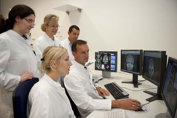
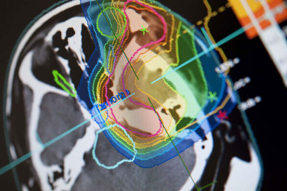
Three-dimensional computer simulation of the tumor area
Radiaion Oncology starts with imaging: Using slice images from computer or magnetic resonance tomography, a three-dimensional computer simulation of the tumor and its surrounding normal tissue is displayed on the monitor within fractions of a second. The physicians then enter certain parameters such as the contours of the tumor and its spatial coordinates, the target dose in the tumor, and the tolerance doses for healthy adjacent tissue. The computer calculates the optimal radiation dose for every single point in the tumor and determines the optimal irradiation direction for the therapy beam. This is known as “three-dimensional computer-based radiation therapy planning”.
Up to 38 individual sessions
The decisive event that leads to the death of a cell is the destruction of its genetic material (DNA). Then, the cell can no longer multiply and dies. The tumor cannot continue to grow. The ion beam must irreparably destroy the genetic string of every single cancer cell. This cannot be done in one session, making several more treatments necessary. This is called fractionation. The breaks between treatments are selected so that healthy tissue affected by the radiation can recover and repair the damage caused by radiation. Cancer cells cannot recover as quickly. The radiation damage thus accumulates in the tumor cells which are destroyed by the radiation treatment.
For most patients, one session lasts only a few minutes. In rare cases, it can last up to 30 minutes. The entire treatment consists of an average of 38 sessions. Several weeks after the radiation, the physicians make a CT or MRI to check whether the tumor has shrunk or even disappeared entirely.
More detailed information on the radiation procedure you´ll find on the Homepage of the Klinik für Radioonkologie und Strahlentherapie.


