


Abdominal and Interventional Imaging Team
Operating at the nexus of interdisciplinary research, Computed Tomography (CT) and Magnetic Resonance Imaging (MRI) hold pivotal roles in diagnosing, planning therapy, and monitoring abdominal diseases. Within this dynamic landscape, the abdominal and interventional imaging research group leads the charge in fundamental and clinical investigations, focusing primarily on the pancreas and liver while transcending boundaries. These initiatives encompass tasks as precise as identifying intrahepatic anatomical structures during liver resection, employing ablative techniques and combination therapies for hepatocellular carcinoma treatment, and pioneering minimally invasive methods for portal vein arterialization.
Embedded within the European Pancreas Center Heidelberg and the Liver Cancer Center Heidelberg, our group's collaborations extend across the Department of General, Visceral and Transplantation Surgery and the Department of Pathology at University Hospital Heidelberg, as well as the Department of Pathology at University Hospital Mainz.
In our pursuit of innovative diagnostics, we assess novel approaches to enhance the (differential-)diagnosis of abdominal diseases, bridging imaging findings with histopathological insights. Moreover, we engineer and validate Artificial Intelligence (AI) algorithms, fostering precise detection and characterization of pancreatic and hepatic lesions.
An integral part of the Body Composition Consortium, our team delves into the predictive significance of body composition parameters (fat, muscle, bone) on patient outcomes within the realm of abdominal diseases. Collaborating closely with abdominal radiologists from esteemed German university hospitals such as Lübeck, Mainz, Berlin, our consortium thrives on collective expertise.
In our work we strive to elevate surgical outcomes, ensure patient safety, and unravel the intricacies of liver and pancreatic diseases.
- Prof. Dr. med. Miriam Klauss
- PD Dr. med. Philipp Mayer
- Dr. med. dr. rer. nat. Caroline Wild
- Dr. med. univ. Chiraz Ben-Salah
- Dr. med. Verena Steinle
- Dr. med. dr. med. Dorottya Móré
- Dr. med. René Mathy
- Andreas Richter
- Parham Tinoush
- Dr. med. univ. Róbert Stollmayer
- M. Sc. Manuel Debic
- Alina Barlemann
- Michelle Güttlein
- Patricia Kupfer
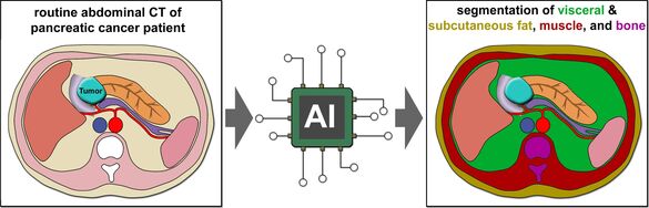
1. CT body-composition analysis using a fully-automated algorithm to predict perioperative complications and prognosis in pancreatic cancer patients.
Verena Steinle, Philipp Mayer
Until lately, radiological body composition analysis necessitated time-consuming manual segmentation of CT or MRI slices. In the present study, we have the opportunity to use a fully automated body composition analysis algorithm which was developed by our cooperation partners from the University Hospital of Essen. The algorithm quantitates body-composition parameters across the whole abdomen. This enables us to perform a large-scale body-composition analysis of preoperative CT scans from pancreatic cancer patients and correlate the results with major perioperative complications and patient survival.
2. CT contrast enhancement to predict prognosis in pancreatic cancer patients
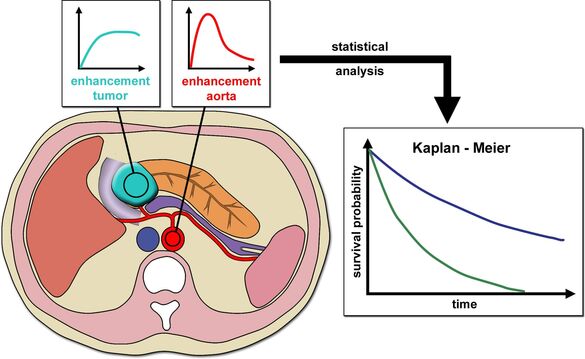
Alina Barlemann, Philipp Mayer
The CT enhancement of pancreatic cancer is thought to be dependent on the histopathological microvessel density, the stromal content, and tumor cell density. All of these parameters were reported to correlate with prognosis of pancreatic cancer patients. Therefore, we hypothesized that CT enhancement of pancreatic cancer lesions can predict patient survival. In the present study, we quantitatively analyze contrast enhancement of treatment-naive pancreatic cancer lesions on multiphase CT scans and correlate the results with patient survival.
3. Prediction of imaging progression of low-risk BD-IPMNs using artificial intelligence
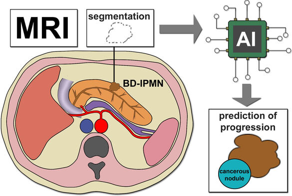
Manuel Debić, Philipp Mayer
Branch duct intraductal papillary mucinous neoplasms (BD-IPMNs) without enhancing solid components/nodules or main duct involvement are considered low-risk lesions. Such patients undergo radiological surveillance to establish the stability of the BD-IPMNs. Most low-risk BD-IPMNs are indolent while some develop features of malignancy during surveillance. Conventional imaging features fail to distinguish BD-IPMNs which will show (imaging) progression, from indolent BD-IPMNs. The present study aims to predict imaging progression of BD-IPMNs using artificial intelligence.
4. Automated LI-RADS classification of liver lesions in gadoxetic acid–enhanced MRI
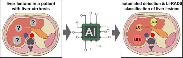
Robert Stollmayer, Philipp Mayer
The Liver Reporting & Data System (LI-RADS) was created by the American College of Radiology to standardize the radiological reporting of liver lesions in patients who are at risk for developing a hepatocellular carcinoma (HCC). Based a diagnostic algorithm, each liver lesion is assigned a LI-RADS category which reflect its probability of being an HCC. Category assignment is time demanding and requires some level of radiological expertise. In the present study, an algorithm for automated detection and LI-RADS classification of lesions on gadoxetic acid–enhanced MRI scans is developed. This algorithm could be useful in the context of tumor boards when there is little time for manual analysis of multiple MRI series as well as for inexperienced radiologists.
5. Imaging differentiation of adenosquamous carcinoma and common pancreatic cancer
Caroline Wild, Philipp Mayer
Adenosquamous carcinoma is a particularly aggressive variant of pancreatic cancer with an inferior prognosis. Most adenosquamous carcinomas are misinterpreted as common pancreatic cancer on initial radiological imaging. The aim of this retrospective study is to enable improved imaging differentiation of adenosquamous carcinoma and common pancreatic cancer using CT and MRI.
6. Is intravenous and rectal contrast administration necessary to diagnose sigmoid diverticulitis?
Dorottya Moré, Philipp Mayer
The current German S3 guideline recommends a CT scan with intravenous (i.v.) and rectal contrast (r.c.) administration for diagnosis of acute sigmoid diverticulitis. There is some evidence from recent studies that a non-contrast (n.c.) CT scan without i.v. or r.c. might be sufficient in this setting. However, studies which compare the diagnostic value of an n.c. scan and an i.v.+r.c. scan in the same patient (paired) are lacking. The present reader study investigates if an n.c. scan is non-inferior to an i.v.+r.c. scan for diagnosis of sigmoid diverticulitis and its complications using a paired analysis.
7. Imaging differentiation of solid pseudopapillary neoplasms and neuroendocrine pancreatic tumors
VAK Moskau Ekatherina Khristenko / Philipp Mayer
Solid pseudopapillary neoplasms are rare pancreatic neoplasms which have a wide epidemiological, clinical, histopathological and radiological overlap with neuroendocrine pancreatic neoplasms. This can lead to misdiagnosis. The present retrospective study aims to identify key CT and MRI features for differentiation of solid pseudopapillary neoplasms and neuroendocrine pancreatic neoplasms.
8. Test-retest variability of diffusion-weighted MRI measurements in pancreatic cancer
Patricia Kupfer / Verena Steinle
Diffusion-weighted imaging (DWI) is part of every pancreatic MRI protocol. Besides the qualitative analysis of DWI which is helpful for lesion detection, quantitative DWI analysis enables characterization of focal pancreatic lesions. However, there is some controversy regarding the test-retest variability and reproducibility of quantitative DWI measurements between scans. The present prospective study aims to investigate the test-retest variability of DWI variables in treatment-naïve pancreatic cancer patients in two consecutive MRI scans (first scan head first, second scan feet first).
9. Detection of local recurrence of pancreatic cancer on dual-energy contrast-enhanced CT
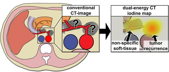
René Mathy / Stephan Skornitzke
In almost all pancreatic cancer patients after tumor resection, non-specific postoperative soft tissue proliferation (PSF) in the area of the operation is observed which is difficult to differentiate from local tumor recurrence using CT-morphological features and/or its location. Differentiation is usually only possible by follow-up CT scans or by PET. In our previous study, we could show that measurement of iodine concentrations in dual-energy contrast-enhanced CT is a valuable parameter for the differentiation of non-specific PSF and local recurrence using a small patient sample from a previous prospective study. In the present study, we aim to confirm these findings in a larger patient cohort.
10. Radio-pathological correlation of MRI features of pancreatic cancer with tumor budding
Philipp Mayer
Tumor budding describes a phenomenon where single cancer cells or small clusters of cancer cells detach from the main tumor mass at the invasive front. It is thought that tumor budding represents the histological basis for local invasion and distant metastases. Tumor budding is negatively correlated with patient survival. In the present study, MRI features of pancreatic cancer are correlated with the histopathological density of tumor buds. Non-invasive prediction of the tumor budding status could enable stratification of pancreatic cancer patients into prognostic subgroups. The study is a cooperation with the Pathological Department of the University Hospital Mainz.
11. Complication and success rate of CT-guided drainage of fluid collections after pancreatic resection
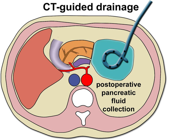
Chiraz Ben-Salah
Fluid collections are a common radiological finding in the postoperative course after pancreatic surgery. Since the fluid collections frequently result from a pancreatic fistula, CT-guided drainages are often inserted into postoperative fluid collections that are not drained by a surgical drainage. The current study aims to retrospectively analyse to complication and success rate of CT-guided drainages of postoperative peripancreatic fluid collections.
12. Detection of cystic pancreatic lesions by MRI in the NAKO study: association with parameters of the metabolic syndrome and pancreatic steatosis
Manuel Debić / Philipp Mayer
This research project is part of the German National Cohort study (NAKO), Germany’s largest population-based study. Women and men aged 20-69 were randomly selected from municipal population registers and recruited as cohort participants. In > 30.000 participants, whole-body MRI scans were conducted.
The present project aims to identify associations and risk factors for the presence of cystic pancreatic lesions (CPLs) on MRI. To date, we have manually analysed the presence, number, size, and location of CPLs on MRI scans of ~11.000 cohort participants and manually segmented the CPLs on T2w-MRIs of a subset of participants. Based on this, we are developing an algorithm for automated detection and segmentation of CPLs which will be applied to the remaining ~20.000 MRI scans. Presence, number, size, and location of CPLs will then be correlated with parameters of the metabolic syndrome and pancreatic steatosis to identify people at risk for presence of CPL.
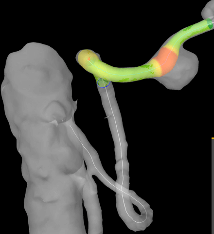
13. DFG Project: Experimental Validation of Fiberoptic Fluorescence Microscopy Technique for Intraoperative Navigation during Liver Resection
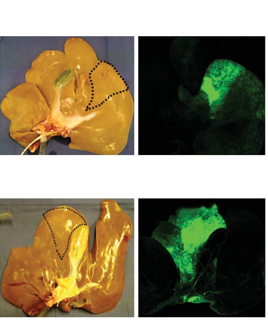
Andreas Richter, Tinoush Parham, De-Hua Chang
This project focuses on enhancing intraoperative navigation during liver resection, a complex operation that demands accurate identification of intrahepatic anatomical structures. To achieve this, we propose a novel approach utilizing Indocyanine Green, Fiberoptic Fluorescence Microscopy, and Macroscopic Imaging for precise resection margin identification. Furthermore, we aim to evaluate the potential benefits of liver segment marking using endothelium-specific antibodies labeled with a liver-specific near-infrared dye. The project will be conducted in three phases, involving (1) ex vivo and (2) in vivo experiments on porcine liver, followed by (3) ex vivo evaluation on human liver parenchyma.
14. The Role of Irreversible Electroporation in the Treatment of Hepatocellular Carcinoma
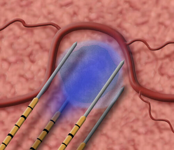
Parham Tinoush, De-Hua Chang
Irreversible electroporation (IRE) is a recent nonthermal technique used for tumor ablation. It works by creating irreversible nanopores in cell membranes, which leads to cell apoptosis. Unlike other ablation methods, IRE is considered safe for tumors located close to large blood vessels and is not affected by the heat-sink effect. Additionally, it has been found to improve the penetration of cytotoxic agents into the surrounding tissue.
Although the combination of IRE with other modalities, such as transarterial chemoembolization (TACE), has shown promising results in experimental studies, clinical evidence supporting its effectiveness is still lacking. Moreover, the specific role of IRE in treating hepatocellular carcinoma (HCC), particularly as a salvage therapy after prior treatments, remains unclear.
This project aims to conduct a retrospective analysis of IRE outcomes and evaluate its effectiveness as a treatment for HCC, both as an independent therapy and as a salvage option. The study will also focus on identifying key factors that impact the short- and long-term results of IRE in HCC patients. By doing so, we hope to gain a better understanding of the potential benefits and limitations of IRE in the context of HCC treatment.
15. THE EMERALD-3 STUDY (NCT05301842): The STRIDE regimen ± lenvatinib given concurrently with TACE compared to TACE alone in patients with HCC amenable to embolisation
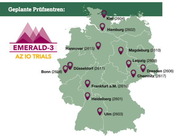
Michael Dill, De-Hua Chang
Intermediate-stage hepatocellular carcinoma (HCC) is a diverse disease with a poor prognosis in the absence of treatment. Transarterial chemoembolization (TACE) is the standard therapy; however, its response is often temporary, and disease progression is common. TACE can prime the HCC microenvironment for immune checkpoint inhibitor (ICI) and anti-VEGF therapy, enhancing anti-tumor responses and increasing VEGF expression. Lenvatinib, a VEGF-receptor kinase inhibitor, has shown improved survival outcomes compared to TACE alone. The STRIDE regimen, consisting of tremelimumab (CTLA-4 inhibitor) and durvalumab (anti-PD-L1 antibody), has demonstrated improved overall survival and manageable safety in unresectable HCC patients. EMERALD-3 aims to evaluate the STRIDE regimen with or without lenvatinib in combination with TACE, building upon previous studies to further advance HCC treatment.
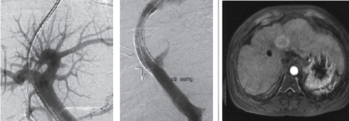
16. Hepatic Stent grafting: Technical success rate, patency rate, peri- and post-interventional complications
Dr. med. René M. Mathy, Daniel Florencia da Ricardo, Prof. D. Chang
Stent graft placement in the hepatic artery is primarily used to treat pseudaneurysms or active bleeding. The advantage of this treatment option is the preservation of the hepatic artery perfusion. The aim of the project is to evaluate the success rate, patency rate and peri- and post-interventional complications as well as relevant influential factors on these parameters. For this purpose, a retrospective analysis of the clinical, laboratory, histopathological, and morphological/interventional radiological data of these patients will be conducted.
17. The diagnosis of hepatocellular carcinoma in patients with transjugular intrahepatic portosystemic shunt: is biopsy mandatory? – a multicentre study
Verena Steinle
To treat complications of portal hypertension in cirrhotic patients, a transjugular intrahepatic portosystemic shunt (TIPS) can be implanted as an artificial channel within the liver that establishes communication between the inflow portal vein and the outflow hepatic vein. The diagnosis of hepatocellular carcinoma is usually made by multiphasic imaging studies demonstrating typical enhancement patterns. Creation of a TIPS often results in characteristic hemodynamic changes of the intrahepatic circulation. The objective of our study is to determine how the presence of TIPS affects the imaging diagnosis of HCC according to the Liver Imaging Reporting and Data System version 2018 (LI-RADSv2018).
18. Analysis and identification of relevant influential factors on success and complications of microwave ablation.
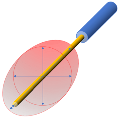
René M. Mathy, De-Hua Chang
Microwave ablation is a thermoablative procedure for the local treatment of tumours. The ablation needle is placed percutaneously and CT-guided in the tumour. This is followed by ablation, in which the surrounding tissue is heated directly by an oscillating electromagnetic field. The aim of this project is to evaluate relevant factors that have an influence on the success rate and recurrence-free survival. Special attention is paid to the analysis of possible influencing factors on the ablation area size.
Selected publications
1. Bao Y, Li JX, Zhou P, Tong Y, Wang LZ, Chang DH, Cai WW, Wen L, Liu J, Xiao YD. Identifying Proliferative Hepatocellular Carcinoma at Pretreatment CT: Implications for Therapeutic Outcomes after Transarterial Chemoembolization. Radiology. 2023 Aug;308(2):e230457.
2. Tong Y, Cai R, Li JX, Chang DH, Wang LZ, Cai WW, Xiao YD. Liver resection versus microwave ablation for hepatocellular carcinoma in ideal candidates for ablation per Barcelona Clinic Liver Cancer staging: a propensity score matching and inverse probability of treatment weighting analysis. Aliment Pharmacol Ther. 2022 Dec;56(11-12):1602-1614.
3. Tan J*, Mathy RM*, Chang DH, Tang T, Zhang ZS, Xiao YD. Combined transarterial iodized oil injection and computed tomography-guided thermal ablation for hepatocellular carcinoma: utility of the iodized oil retention pattern. Abdom Radiol (NY). 2022 Jan;47(1):431-442. *Contributed equally
4. Mohr I, Vogeler M, Pfeiffenberger J, Sprengel SD, Klauss M, Radeleff B, Teufel A, Chang DH, Springfeld C, Longerich T, Merle U, Mehrabi A, Weiss KH, Mieth M. Clinical effects and safety of different transarterial chemoembolization methods for bridging and palliative treatments in hepatocellular carcinoma. J Cancer Res Clin Oncol. 2022 Nov;148(11):3163-3174.
5. Ekaterina Khristenko, Thomas Hank, Matthias M. Gaida, Hans‑Ulrich Kauczor, Thilo Hackert, Miriam Klauß, Philipp Mayer. Imaging features of intraductal tubulopapillary neoplasm of the pancreas and its differentiation from conventional pancreatic ductal adenocarcinoma. Sci Rep. 2022 Sep 16;12(1):15557.
6. Philipp Mayer, Athanasios Giannakis, Miriam Klauß, Matthias M Gaida, Frank Bergmann, Hans-Ulrich Kauczor, Manuel Feisst, Thilo Hackert, Martin Loos. Radiological evaluation of pancreatic cancer: What is the significance of arterial encasement >180° after neoadjuvant treatment? Eur J Radiol. 2021 Apr;137:109603.
7. Philipp Mayer, Franziska Fritz, Marco Koell, Stephan Skornitzke, Frank Bergmann, Matthias M. Gaida, Thilo Hackert, Klaus Maier-Hein, Frederik B. Laun, Hans-Ulrich Kauczor, Lars Grenacher, Miriam Klauß and Wolfram Stiller. Assessment of tissue perfusion of pancreatic cancer as potential imaging biomarker by means of Intravoxel incoherent motion MRI and CT perfusion: correlation with histological microvessel density as ground truth. Cancer Imaging. 2021 Jan 19;21(1):13.
8. Mathy RM, Tinoush P, da Florencia RD, Braun A, Ghamarnejad O, Radeleff B, Kauczor HU, Chang DH. Impact of needle positioning on ablation success of irreversible electroporation: a unicentric retrospective analysis. Sci Rep. 2020 Dec 14;10(1):21902.
9. Philipp Mayer, Christine Dinkic, Ralf Jesenofsky, Miriam Klauß, Peter Schirmacher, Ulrike Dapunt, Thilo Hackert, Florian Uhle, G. Maria Hänsch, Matthias M. Gaida. Changes in the microarchitecture of the pancreatic cancer stroma are linked to neutrophil-dependent reprogramming of stellate cells and reflected by diffusion-weighted magnetic resonance imaging. Theranostics. 2018 Jan 1;8(1):13-30.
10. Miriam Klauß, Philipp Mayer, Frank Bergmann, Klaus Maier-Hein, Judith Hase, Thilo Hackert, Hans-Ulrich Kauczor, Lars Grenacher, Bram Stieltjes. Correlation of Histological Vessel Characteristics and Diffusion-Weighted Imaging Intravoxel Incoherent Motion-Derived Parameters in Pancreatic Ductal Adenocarcinomas and Pancreatic Neuroendocrine Tumors. Invest Radiol. 2015 Nov;50(11):792-7.