


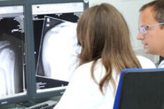

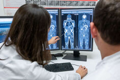
Lung and Cardiovascular Imaging Team
Computed tomography (CT) still remains the primary radiological method for diagnosing lung disorders such as chronic obstructive pulmonary disease (COPD), interstitial lung disease (ILD) and bronchial carcinoma. By identifying radiological biomarkers, the group could contribute to a better understanding of these diseases. Moreover, in the last years, more and more possibilities are opening up to assess the properties of tumors (malignancy, response to therapy) and to investigate the function of the lungs and airways. Moreover, the introduction of quantitative software tools contributed to an objective assessment of lung and airway abnormalities. To reduce the radiation exposure of CT examinations of the chest, low-dose CT technologies and magnetic resonance imaging (MRI) of the lungs were developed. Essential components of the lung MRI technology were discovered and refined in Heidelberg. Certain lung abnormalities can now be visualized with a similar sensitivity than in CT. Especially for radiation-sensitive patients such as pregnant women and children, MRI can already replace CT in many cases. As a radiation-free technology, it helps the Heidelberg researchers to gain essential knowledge about diseases of the respiratory tract such as cystic fibrosis (CF), bronchial asthma, chronic bronchitis or COPD and thus contributes to a better understanding of these diseases and the emergence of new, more efficient treatment methods. Due to the possibility of dynamic measurements, important functional parameters such as perfusion deficits can also be revealed with MRI.
The primary research concern of the doctors and scientists of the group "Lung Imaging Group", is the development and application of new radiological methods to examine structural and functional lung changes in patients with common congenital or acquired chronic lung diseases. Therefore, the group works closely and intensively with the specialists of the Thoraxklinik (Head of Radiology: Prof. Heussel) and the German Cancer Research Center (dkfz). Scientists of the group are members of the Platform Imaging of the German Center for Lung Research (DZL) and the Translational Lung Research Center (TLRC) in Heidelberg.
- PD Dr. med. Katharina Abbasi Dezfouli
- Prof. Dr. med. Hans-Ulrich Kauczor
- Prof. Dr. med. Jürgen Biederer
- PD Dr. med. Philip Konietzke
- Dr. rer. nat. Oliver Weinheimer
- PD Dr. rer. nat. Simon Triphan
- Dr. med. Claudius Melzig
- Dr. med. Oliver Sedlaczek
- Dr. med. Patricia Leutz-Schmidt
- Dr. med. Viktoria Palm
- PD Dr. med. Lena Wucherpfennig
- Dr. med. Sebastian Nauck
- Dr. med. Arved Bischoff
- Dr. med. Marvin Eissler
- Dr. med. Antonia Maletzko
- Carley Stewart, Ph.D.
- Dr. Bettina Budai
- Sonja Gestewitz
- Johannes Dunsche
- Tamas Fero
PhD Students:
- M. Sc. Julian Grolig
- M. Sc. Sarah Muller
- M. Sc. Yiling Xu
- M. Sc. Shengkai Zhao
- M. Sc. Christian Heidt
- Claudia Benke
- Johanna Thomä
- Johanna K. Z. Becker
Research Projects
1. Magnetic resonance imaging (MRI) of the lungs
Mark Wielpütz, Lena Wucherpfennig, Simon Triphan, Jürgen Biederer, Johanna Becker
Magnetic resonance imaging (MRI) of the lungs is technically challenging due to the low proton density of the lung parenchyma and the rapid signal decay. Nevertheless, standard protocols, which enable the depiction of subtle morphological lung and airway pathologies with high diagnostic accuracy, have been established in the last years. In particular, diseases that are associated with an increase in fluid and tissue and thus with an increase in protons, such as bronchial wall thickening, mucus plugging and infiltrates, can be easily visualized by MRI. The group's work has already shown that manifestations of CF lung disease can be identified using MRI even before the onset of symptoms in infants and toddlers and thus led to an earlier initiation of therapy in the pre-symptomatic stage, before irreversible lung damage occurs.
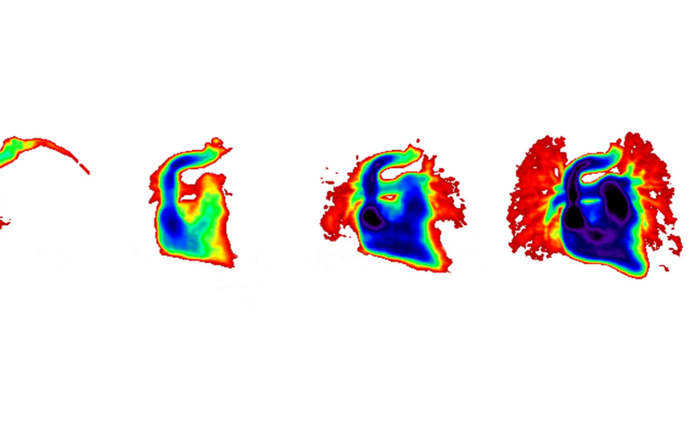
In addition to morphological imaging, MRI offers the possibility of additional functional information. Using dynamic MRI sequences, respiratory mechanics, lung ventilation and lung perfusion can be determined. These functional parameters enable conclusions to be drawn about the pathophysiology of the lungs and improve the imaging of diseases such as COPD, CF and pulmonary embolism. The combination of morphological and functional lung imaging with high spatial and temporal resolution is a major advantage of MRI compared to other imaging modalities. For example, approaches for quantifying perfusion defects were developed in this research group.
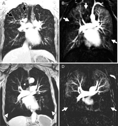
High-resolution MR angiography can also provide information about the pulmonary arteries and the bronchial arteries, which are often enlarged in chronic lung diseases.
In addition, as a radiation-free, non-invasive examination modality, MRI offers the possibility of repeated examinations, so that disease progression or therapeutic response e.g. to chemotherapy in patients with bronchial carcinoma or to gene modulator therapies in patients with CF can be evaluated.
Because of these advantages, MRI of the lungs offers a broad research field for the study group.
Publications
- Bayfield KJ, Weinheimer O, Boyton C, Fitzpatrick R, Middleton A, Kennedy B, Blaxland A, Jayasuriya G, Caplain N, Issa H, Goetti R, Wielpütz MO, Yu L, Galban CJ, Robinson TE, Bartholmai B, Fitzgerald D, Selvadurai H, Robinson PD. Implementation and evaluation of ultra-low dose CT in early cystic fibrosis lung disease. Eur Respir J. 2023 Jul 7;62(1):2300286. doi: 10.1183/13993003.00286-2023.
- Wucherpfennig L, Triphan SMF, Wege S, Kauczor HU, Heussel CP, Sommerburg O, Stahl M, Mall MA, Eichinger M, Wielpütz MO. Elexacaftor/Tezacaftor/Ivacaftor Improves Bronchial Artery Dilatation Detected by Magnetic Resonance Imaging in Patients with Cystic Fibrosis. Ann Am Thorac Soc. 2023 Nov;20(11):1595-1604. doi: 10.1513/AnnalsATS.202302-168OC.
- Wucherpfennig L, Wuennemann F, Eichinger M, Seitz A, Baumann I, Stahl M, Graeber SY, Zhao S, Chung J, Schenk JP, Alrajab A, Kauczor HU, Mall MA, Sommerburg O, Wielpütz MO. Long-term effects of lumacaftor/ivacaftor on paranasal sinus abnormalities in children with cystic fibrosis detected with magnetic resonance imaging. Front Pharmacol. 2023 Apr 10;14:1161891. doi: 10.3389/fphar.2023.1161891.
- Muller, S.M, Ram S, Bayfield KJ, Gestewitz S, Yu Lifeng, Wielpütz MO, Kauczor HU, Heussel CP, Robinson TE, Bartholmai, BJ, Hatt CR, Robinson PD, Galban CJ & Weinheimer O. Deep Learning-Based Air Trapping Quantification Using Paired Inspiratory-Expiratory Ultra-low Dose CT. Medical Image Computing and Computer Assisted Intervention – MICCAI 2023. MICCAI 2023. Lecture Notes in Computer Science, vol 14222. Springer, Cham. 2023. doi: 10.1007/978-3-031-43898-1_42
- Wucherpfennig L, Wuennemann F, Eichinger M, Schmitt N, Seitz A, Baumann I, Stahl M, Graeber SY, Chung J, Schenk JP, Alrajab A, Kauczor HU, Mall MA, Sommerburg O, Wielpütz MO. Longitudinal Magnetic Resonance Imaging Detects Onset and Progression of Chronic Rhinosinusitis from Infancy to School Age in Cystic Fibrosis. Ann Am Thorac Soc. 2023 May;20(5):687-697. doi: 10.1513/AnnalsATS.202209-763OC.
- Palm V, Norajitra T, von Stackelberg O, Heussel CP, Skornitzke S, Weinheimer O, Kopytova T, Klein A, Almeida SD, Baumgartner M, Bounias D, Scherer J, Kades K, Gao H, Jäger P, Nolden M, Tong E, Eckl K, Nattenmüller J, Nonnenmacher T, Naas O, Reuter J, Bischoff A, Kroschke J, Rengier F, Schlamp K, Debic M, Kauczor HU, Maier-Hein K, Wielpütz MO. AI-Supported Comprehensive Detection and Quantification of Biomarkers of Subclinical Widespread Diseases at Chest CT for Preventive Medicine. Healthcare (Basel). 2022 Oct 29;10(11):2166. doi: 10.3390/healthcare10112166.
- Wucherpfennig L, Triphan SMF, Wege S, Kauczor HU, Heussel CP, Schmitt N, Wuennemann F, Mayer VL, Sommerburg O, Mall MA, Eichinger M, Wielpütz MO. Magnetic resonance imaging detects improvements of pulmonary and paranasal sinus abnormalities in response to elexacaftor/tezacaftor/ivacaftor therapy in adults with cystic fibrosis. J Cyst Fibros. 2022 Nov;21(6):1053-1060. doi: 10.1016/j.jcf.2022.03.011.
- Do TD, Skornitzke S, Merle U, Kittel M, Hofbaur S, Melzig C, Kauczor HU, Wielpütz MO, Weinheimer O. COVID-19 pneumonia: Prediction of patient outcome by CT-based quantitative lung parenchyma analysis combined with laboratory parameters. PLoS One. 2022 Jul 29;17(7):e0271787. doi: 10.1371/journal.pone.0271787.
- Skornitzke S, Vats N, Kopytova T, Tong EWY, Hofbauer T, Weber TF, Rehnitz C, von Stackelberg O, Maier-Hein K, Stiller W, Biederer J, Kauczor HU, Heußel CP, Wielpütz M, Palm V. Asynchronous calibration of quantitative computed tomography bone mineral density assessment for opportunistic osteoporosis screening: phantom-based validation and parameter influence evaluation. Sci Rep. 2022 Dec 1;12(1):20729. doi: 10.1038/s41598-022-24546-2.
- Triphan SMF, Weinheimer O, Gutberlet M, Heußel CP, Vogel-Claussen J, Herth F, Vogelmeier CF, Jörres RA, Kauczor HU, Wielpütz MO, Biederer J, Jobst BJ; COSYCONET Study Group. Echo Time-Dependent Observed Lung T1 in Patients With Chronic Obstructive Pulmonary Disease in Correlation With Quantitative Imaging and Clinical Indices. J Magn Reson Imaging. 2021 Nov;54(5):1562-1571. doi: 10.1002/jmri.27746.
- Stahl M, Steinke E, Graeber SY, Joachim C, Seitz C, Kauczor HU, Eichinger M, Hämmerling S, Sommerburg O, Wielpütz MO, Mall MA. Magnetic Resonance Imaging Detects Progression of Lung Disease and Impact of Newborn Screening in Preschool Children with Cystic Fibrosis. Am J Respir Crit Care Med. 2021 Oct 15;204(8):943-953. doi: 10.1164/rccm.202102-0278OC. PMID: 34283704.
- Palm V, Rengier F, Rajiah P, Heussel CP, Partovi S. Acute Pulmonary Embolism: Imaging Techniques, Findings, Endovascular Treatment and Differential Diagnoses. Rofo. 2020; 192(01): 38-49. doi: 10.1055/a-0900-4200
- Triphan SMF, Stahl M, Jobst BJ, Sommerburg O, Kauczor HU, Schenk JP, Alrajab A, Eichinger M, Mall MA, Wielpütz MO. Echo Time-Dependence of Observed Lung T1 in Patients With Cystic Fibrosis and Correlation With Clinical Metrics. J Magn Reson Imaging. 2020 Dec;52(6):1645-1654. doi: 10.1002/jmri.27271.
- Konietzke P, Wielpütz MO, Wagner WL, Wuennemann F, Kauczor HU, Heussel CP, Eichinger M, Eberhardt R, Gompelmann D, Weinheimer O. Quantitative CT detects progression in COPD patients with severe emphysema in a 3-month interval. Eur Radiol. 2020 May;30(5):2502-2512. doi: 10.1007/s00330-019-06577-y.
- Wielpütz MO, Eichinger M, Wege S, Eberhardt R, Mall MA, Kauczor HU, Puderbach MU, Risse F, Heußel CP, Heußel G. Midterm Reproducibility of Chest Magnetic Resonance Imaging in Adults with Clinically Stable Cystic Fibrosis and Chronic Obstructive Pulmonary Disease. Am J Respir Crit Care Med. 2019 Jul 1;200(1):103-107. doi: 10.1164/rccm.201812-2356LE.
- Jobst BJ, Weinheimer O, Buschulte T, Trauth M, Tremper J, Delorme S, Becker N, Motsch E, Groß ML, Trotter A, Eichinger M, Kauczor HU, Wielpütz MO. Longitudinal airway remodeling in active and past smokers in a lung cancer screening population. Eur Radiol. 2019 Jun;29(6):2968-2980. doi: 10.1007/s00330-018-5890-4.
- Palm V, Sheng R, Mayer P, Weiss KH, Springfeld C, Mehrabi A, Longerich T, Berger AK, Kauczor HU, Weber TF. Imaging features of fibrolamellar hepatocellular carcinoma in gadoxetic acid-enhanced MRI. Cancer Imaging. 2018 Mar 1;18(1):9. doi: 10.1186/s40644-018-0143-y.
- Stahl M, Wielpütz MO, Graeber SY, Joachim C, Sommerburg O, Kauczor HU, Puderbach M, Eichinger M, Mall MA. Comparison of Lung Clearance Index and Magnetic Resonance Imaging for Assessment of Lung Disease in Children with Cystic Fibrosis. Am J Respir Crit Care Med. 2017 Feb 1;195(3):349-359. doi: 10.1164/rccm.201604-0893OC.
- Triphan SM, Jobst BJ, Breuer FA, Wielpütz MO, Kauczor HU, Biederer J, Jakob PM. Echo time dependence of observed T1 in the human lung. J Magn Reson Imaging. 2015 Sep;42(3):610-6. doi: 10.1002/jmri.24840.
- Triphan SM, Breuer FA, Gensler D, Kauczor HU, Jakob PM. Oxygen enhanced lung MRI by simultaneous measurement of T1 and T2 * during free breathing using ultrashort TE. J Magn Reson Imaging. 2015 Jun;41(6):1708-14. doi: 10.1002/jmri.24692.
- Wielpütz MO, Puderbach M, Kopp-Schneider A, Stahl M, Fritzsching E, Sommerburg O, Ley S, Sumkauskaite M, Biederer J, Kauczor HU, Eichinger M, Mall MA. Magnetic resonance imaging detects changes in structure and perfusion, and response to therapy in early cystic fibrosis lung disease. Am J Respir Crit Care Med. 2014 Apr 15;189(8):956-65. doi: 10.1164/rccm.201309-1659OC.
- Weinheimer O, Achenbach T, Bletz C, Duber C, Kauczor HU, Heussel CP. About objective 3-d analysis of airway geometry in computerized tomography. IEEE Trans Med Imaging. 2008 Jan;27(1):64-74. doi: 10.1109/TMI.2007.902798.