
![[Translate to English:]](/fileadmin/_processed_/2/3/csm_20210614_MedKlinik_004_26a0b1fc8e.jpg "[Translate to English:]")
![[Translate to English:]](/fileadmin/_processed_/1/c/csm_Patientenbetreuung_b795e65d27.jpg "[Translate to English:]")

![[Translate to English:]](/fileadmin/_processed_/a/3/csm_I_202008040096588_7d1e8a84d4.jpg "[Translate to English:]")
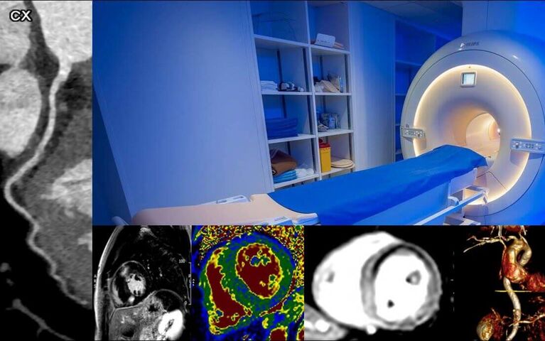
Cardiovascular Imaging (cCTA & CMR)
Welcome to the homepage of the division for cardiac imaging at the Heidelberg University Hospital
Our division for cardiac imaging includes the modalities cardiovascular magnetic resonance imaging (CMR) and coronary computed tomography angiography (cCTA). It is supervised by our working group Cardiovascular Imaging (CT/MRI) of the Department of Cardiology, Angiology, and Pneumology.
Cardiovascular magnetic resonance imaging (CMR)
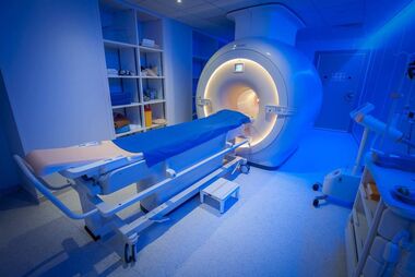
Cardiovascular magnetic resonance imaging (CMR) has undergone rapid development in recent years and plays an increasingly important role in the diagnosis, treatment planning, and risk assessment of patients with heart diseases. In our department, we have been conducting CMR examinations since 2006, having performed over 40,000 examinations to date. Currently, with generous support from the Dietmar Hopp Foundation, we have two modern MR devices (1.5 and 3T) dedicated exclusively to the performance of CMRs in specially adapted facilities.
The 1.5 Tesla device allows for the examination of patients with the most common implantable cardiac devices, while the 3 Tesla device, due to its larger opening, enables the examination of overweight patients and those with claustrophobia.
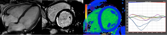
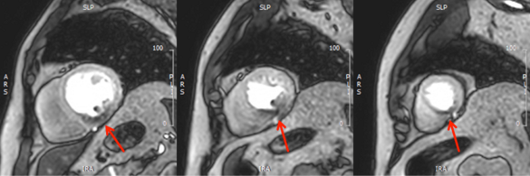
Due to its high spatial and temporal resolution as well as excellent tissue depiction, CMR allows for the excellent and accurate visualization and characterization of the composition and function of all heart and vascular components. As a non-invasive procedure that does not involve ionizing radiation (X-rays), CMR is also an extremely safe method, allowing for multiple follow-up examinations, such as in the case of myocardial diseases. Furthermore, it offers the advantage over other imaging modalities of being able to non-invasively characterize the tissue of the heart, which would otherwise only be possible through histological analysis after biopsy. This "tissue characterization" is done through special late gadolinium enhancement images, where patients receive a well-tolerated MRI contrast agent, and through modern mapping techniques, which mostly do not require contrast agent application. These techniques can detect fibrosis, scars (e.g., after a heart attack), or water retention (edema) in the heart muscle.
Thanks to continuous technical and scientific advancements, CMR can now provide targeted answers to a variety of specific questions. CMR holds significant importance in the ischemia testing in coronary heart disease. Through pharmacological stress tests using vasodilators (adenosine) or positive inotropic agents (dobutamine), perfusion or resulting motion abnormalities of the heart muscle can be identified. This method allows for the accurate detection of hemodynamically relevant stenoses in the coronary arteries and determines their impact on myocardial perfusion with high precision.
Another important field is the diagnosis and risk stratification of heart muscle diseases (cardiomyopathies). In addition to assessing the function and structure of the heart, tissue characterization is of outstanding importance. The identification of scarring and fibrosis leads to a better understanding of the underlying etiology and can predict individual risk for adverse cardiac events.
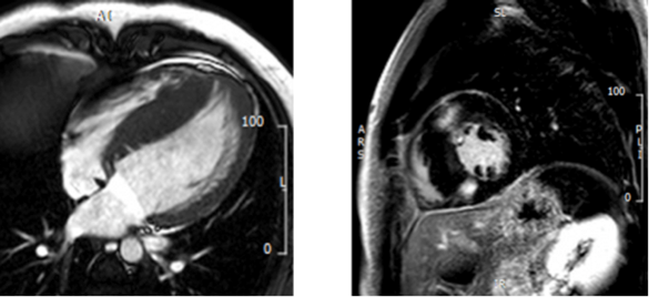
Further significant focuses of CMR include the detection of inflammatory changes in the heart muscle (e.g., myocarditis), differentiation of storage diseases of the heart (e.g., amyloidosis), monitoring of heart function during chemotherapy, and assessment of complex congenital heart defects. Nowadays, CMR can also precisely measure heart valve defects and intracardiac shunts.
In collaboration with the Department of Pediatric Cardiology and Congenital Heart Defects, we also conduct prenatal cardiac MRI examinations on unborn children (fetal cardiac MRI / fetal CMR). This allows us to significantly improve the assessment of the severity of heart defects in cases of unclear diagnoses. For more information and contact information click here (Link).
Coronary Computed Tomography Angiography (cCTA)
Coronary Computed Tomography Angiography (cCTA)
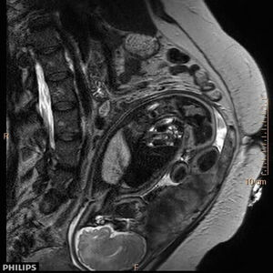
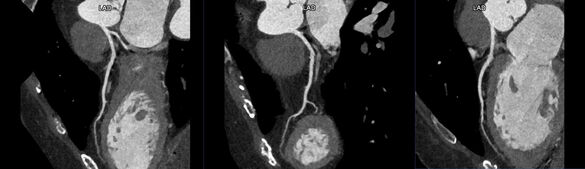
The anatomical visualization of the coronary arteries and the major vessels near the heart is an integral part of cardiological diagnosis and crucial for treatment planning in patients with chest pain. While invasive coronary angiography was once the only method to examine the coronary arteries, cCTA is now established with the same diagnostic reliability and is globally recommended by the current guidelines.
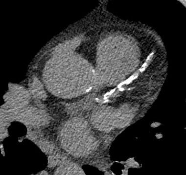
Alternatively or in addition calcifications in the coronary arteries can be assessed under low radiation exposure and without the use of contrast agents with the so-called calcium scoring.
In addition to assess stable chest pain, cCTA is also valuable in an emergency setting. In a single contrast-enhanced CT examination, it can reliably exclude not only stenoses in the coronary arteries but also other potentially life-threatening conditions such as pulmonary artery embolism or a disease of the main artery (e.g., aortic dissection). Another increasingly important field, driven by the technical progress of invasive cardiology, is CT angiography to plan complex interventional heart valve procedures (e.g., prior to transcatheter aortic valve implantation).
In collaboration with the Department of Diagnostic and Interventional Radiology, we conduct outpatient and inpatient cardiac CT examinations using two state-of-the-art CT devices (Philips IQon Spectral CT and Siemens Somatom Force). We are a certified center of the German Society of Cardiology and have decades of experience in conducting these examinations.
Members
![[Translate to English:] Portrait von Dr. med. Florian André](/fileadmin/_processed_/9/a/csm_andre_florian203_1_b2d2849f97.jpg "[Translate to English:] Portrait von Dr. med. Florian André")

![[Translate to English:] Portrait von PD Dr. med. Dirk Loßnitzer, FESC](/fileadmin/_processed_/9/b/csm_Lossnitzer-1_1420441517.jpg "[Translate to English:] Portrait von PD Dr. med. Dirk Loßnitzer, FESC")
![[Translate to English:] Portrait von Dr. med. Ailis Ceara Haney](/fileadmin/_processed_/e/2/csm_Haney__Ailis_Med3_05_c987f3e7de.jpg "[Translate to English:] Portrait von Dr. med. Ailis Ceara Haney")
![[Translate to English:] Portrait von Andreas Ochs](/fileadmin/_processed_/1/0/csm_Ochs_Andreas_4725b3ed79.jpg "[Translate to English:] Portrait von Andreas Ochs")


Daniel Asmussen
Senior medical technician for functional diagnostics
(Research Group Cardiovascular Imaging)

Katharina Bösenberg
Medical technician for functional diagnostics
(Research Group Cardiovascular Imaging)
Research Funding
Selected publications
Haney A, Salatzki J, Hund H, Friedrich M, Giannitsis E, Frey N, Steen H, Lossnitzer D, Riffel J, André F. Prognostic value of negative stress cardiac magnetic resonance imaging in patients with moderate-severe coronary artery stenosis. Frontiers in Cardiovascular Medicine. 2023. 10. 10.3389/fcvm.2023.1264374.<o:p></o:p>
Weberling LD, Seitz S, Salatzki J, Ochs A, Heins J, Haney AC, Siry D, Frey N, André F, Steen H. Safety of dobutamine or adenosine stress cardiac magnetic resonance imaging in patients with left ventricular thrombus. Clin Res Cardiol. 2023 Oct 16. doi: 10.1007/s00392-023-02317-x. Epub ahead of print. PMID: 37843560.<o:p></o:p>
Andre F, Seitz S, Fortner P, Allmendinger T, Sommer A, Brado M, Sokiranski R, Fink J, Kauczor HU, Heussel CP, Herth F, Frey N, Görich J, Buss SJ. Simultaneous assessment of heart and lungs with gated high-pitch ultra-low dose chest CT using artificial intelligence-based calcium scoring. Eur J Radiol Open. 2023 Feb 15;10:100481. doi: 10.1016/j.ejro.2023.100481. PMID: 36852255; PMCID: PMC9958356.<o:p></o:p>
Salatzki J, Ochs A, Kirchgäßner N, Heins J, Seitz S, Hund H, Mereles D, Friedrich MG, Katus HA, Frey N, André F, Ochs MM. Safety of Stress Cardiac Magnetic Resonance in Patients With Moderate to Severe Aortic Valve Stenosis. J Cardiovasc Imaging. 2023 Jan;31(1):26-38. doi: 10.4250/jcvi.2022.0063. PMID: 36693342; PMCID: PMC9880345.<o:p></o:p>
Weberling LD, Seitz S, Salatzki J, Ochs A, Haney AC, Siry D, Heins J, Steen H, Frey N, André F. Safety, accuracy, and prediction of prognosis in patients with end-stage chronic kidney disease undergoing dobutamine stress cardiac magnetic resonance imaging. Front Cardiovasc Med. 2023 Aug 30;10:1228691. doi: 10.3389/fcvm.2023.1228691. PMID: 37711564; PMCID: PMC10498284.<o:p></o:p>
Weberling LD, Lossnitzer D, Frey N, André F. Coronary Computed Tomography vs. Cardiac Magnetic Resonance Imaging in the Evaluation of Coronary Artery Disease. Diagnostics (Basel). 2022 Dec 30;13(1):125. doi: 10.3390/diagnostics13010125. PMID: 36611417; PMCID: PMC9818886.<o:p></o:p>
Siry D, Riffel J, Salatzki J, André F, Weberling LD, Ochs M, Atia NA, Hillier E, Albert D, Katus HA, Giannitsis E, Frey N, Friedrich MG. A head-to-head comparison of fast-SENC and feature tracking to LV long axis strain for assessment of myocardial deformation in chest pain patients. BMC Med Imaging. 2022 Sep 5;22(1):159. doi: 10.1186/s12880-022-00886-3. PMID: 36064332; PMCID: PMC9442977.<o:p></o:p>
Salatzki J, Giannitsis E, Hegenbarth A, Mueller-Hennessen M, André F, Katus HA, Frey N, Biener M. Correlation of serial high-sensitivity cardiac Troponin T values to infarct mass determined by cardiac magnetic resonance imaging: a validation study. Eur Heart J Acute Cardiovasc Care. 2022 Nov 30;11(11):826-833. doi: 10.1093/ehjacc/zuac122. PMID: 36184989.<o:p></o:p>
Weberling LD, Steen H, Frey N, André F. Large Mobile Left Ventricular Thrombi Formation in a 32-Year-Old Despite Direct Oral Anticoagulation With Dabigatran. JACC Case Rep. 2022 Aug 17;4(16):1015-1019. doi: 10.1016/j.jaccas.2022.05.033. PMID: 36062055; PMCID: PMC9434649.<o:p></o:p>
Siry D, Riffel JH, Salatzki J, Andre F, Ochs M, Weberling LD, Giannitsis E, Katus HA, Friedrich MG. Hypverventilation strain CMR imaging in patients with acute chest pain. Sci Rep. 2022 Aug 9;12(1):13584. doi: 10.1038/s41598-022-17856-y. PMID: 35945332; PMCID: PMC9363440.<o:p></o:p>
Weberling LD, Friedrich MG. Sauerstoffsensitive kardiale Magnetresonanztomographie [Oxygenation-sensitive cardiac magnetic resonance imaging]. Radiologie (Heidelb). 2022 Nov;62(11):971-976. German. doi: 10.1007/s00117-022-01049-9. Epub 2022 Jul 29. PMID: 35904573.<o:p></o:p>
Ochs A, Nippes M, Salatzki J, Weberling LD, Riffel J, Müller-Hennessen M, Giannitsis E, Osman N, Stehning C, André F, Katus HA, Frey N, Friedrich MG, Ochs MM. Dynamic Handgrip Exercise: Feasibility and Physiologic Stress Response of a Potential Needle-Free Cardiac Magnetic Resonance Stress Test. Front Cardiovasc Med. 2021 Nov 29;8:755759. doi: 10.3389/fcvm.2021.755759. PMID: 34912862; PMCID: PMC8666587.<o:p></o:p>
Salatzki J, Fischer T, Riffel J, André F, Hirschberg K, Ochs A, Hund H, Müller-Hennessen M, Giannitsis E, Friedrich MG, Scholz E, Frey N, Katus HA, Ochs M. Presence of contractile impairment appears crucial for structural remodeling in idiopathic left bundle-branch block. J Cardiovasc Magn Reson. 2021 Apr 1;23(1):39. doi: 10.1186/s12968-021-00731-6. PMID: 33789682; PMCID: PMC8015193.<o:p></o:p>
Ochs A, Riffel J, Ochs MM, Arenja N, Fritz T, Galuschky C, Schuster A, Bruder O, Mahrholdt H, Giannitsis E, Frey N, Katus HA, Buss SJ, André F. Myocardial mechanics in dilated cardiomyopathy: prognostic value of left ventricular torsion and strain. J Cardiovasc Magn Reson. 2021 Dec 2;23(1):136. doi: 10.1186/s12968-021-00829-x. PMID: 34852822; PMCID: PMC8638178.<o:p></o:p>
Salatzki J, Mohr I, Heins J, Cerci MH, Ochs A, Paul O, Riffel J, André F, Hirschberg K, Müller-Hennessen M, Giannitsis E, Friedrich MG, Merle U, Weiss KH, Katus HA, Ochs M. The impact of Wilson disease on myocardial tissue and function: a cardiovascular magnetic resonance study. J Cardiovasc Magn Reson. 2021 Jun 24;23(1):84. doi: 10.1186/s12968-021-00760-1. PMID: 34162411; PMCID: PMC8223377.<o:p></o:p>
Steen H, Montenbruck M, Kelle S, Esch S, Schwarz AK, Giusca S, Korosoglou G. Fast-Strain Encoded Cardiac Magnetic Resonance During Vasodilator Perfusion Stress Testing. Front Cardiovasc Med. 2021 Nov 17;8:765961. doi: 10.3389/fcvm.2021.765961. PMID: 34869679; PMCID: PMC8635645.<o:p></o:p>
Giusca S, Korosoglou G, Montenbruck M, Geršak B, Schwarz AK, Esch S, Kelle S, Wülfing P, Dent S, Lenihan D, Steen H. Multiparametric Early Detection and Prediction of Cardiotoxicity Using Myocardial Strain, T1 and T2 Mapping, and Biochemical Markers: A Longitudinal Cardiac Resonance Imaging Study During 2 Years of Follow-Up. Circ Cardiovasc Imaging. 2021 Jun;14(6):e012459. doi: 10.1161/CIRCIMAGING.121.012459. Epub 2021 Jun 15. PMID: 34126756; PMCID: PMC8208092.<o:p></o:p>
Steen H, Giusca S, Montenbruck M, Patel AR, Pieske B, Florian A, Erley J, Kelle S, Korosoglou G. Left and right ventricular strain using fast strain-encoded cardiovascular magnetic resonance for the diagnostic classification of patients with chronic non-ischemic heart failure due to dilated, hypertrophic cardiomyopathy or cardiac amyloidosis. J Cardiovasc Magn Reson. 2021 Apr 5;23(1):45. doi: 10.1186/s12968-021-00711-w. PMID: 33823860; PMCID: PMC8025329.<o:p></o:p>
Korosoglou G, Giusca S, Montenbruck M, Patel AR, Lapinskas T, Götze C, Zieschang V, Al-Tabatabaee S, Pieske B, Florian A, Erley J, Katus HA, Kelle S, Steen H. Fast Strain-Encoded Cardiac Magnetic Resonance for Diagnostic Classification and Risk Stratification of Heart Failure Patients. JACC Cardiovasc Imaging. 2021 Jun;14(6):1177-1188. doi: 10.1016/j.jcmg.2020.10.024. Epub 2021 Jan 13. PMID: 33454266.<o:p></o:p>