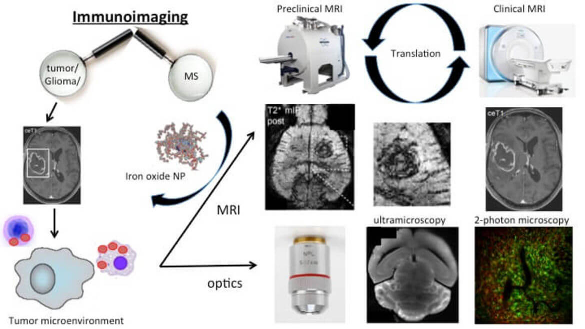
Sektion Immuno-Imaging
Die Sektion Immuno-Imaging verfolgt in enger Zusammenarbeit mit der Klinischen Kooperationseinheit Neuroimmunologie und Hirntumorimmunologie (DKFZ, Leiter Prof. Platten, https://www.dkfz.de/de/neuroimmunologie/Immunoimaging-Team.html) das Ziel immunologische Vorgänge in Autoimmunität (Multiple Sklerose, MS) und Neuroonkologie (Hirntumoren) zu visualisieren. Dazu nutzen wir molekulares und zelluläres Imaging mittels Hochfeld-MRT und korrelierter optischer Bildgebung (Ultramikroskopie/Clearing, 2-Photonen Mikroskopie, Konfokalmikroskopie). Ziel ist es Immunzelldynamiken und deren Änderungen unter (Immun-) Therapien im neuroinflammatorischen Kontext zu visualisieren und zu quantifizieren. Dazu werden neue immuntherapeutische Verfahren (Checkpoint-Inhibition, Vakzine, Adoptiver T-Zell Transfer gegen Tumor-Neoepitope) in präklinischen Gliom-Modellen getestet, um Imagingmarker für adaptive und angeborene Immunzellreaktionen zu entwickeln. Das Immunzelltracking erfolgt dabei mittels Eisen-Nanopartikeln und multiparametrischer MRT.
Ein weiterer Schwerpunkt liegt auf neuen Erkrankungsmodellen zur Darstellung von Tumorzellinfiltration und deren Korrelaten in MRT und optischer Bildgebung (Kooperation Prof. Winkler).
Zudem erfolgen klinische Studien in MS und Hirntumorpatienten, um präklinische Imaging Biomarker in die klinische Praxis zu überführen.
Ziel ist schlussendlich die Etablierung von Imaging-Biomarkern, die die Charakterisierung des entzündlichen Mikromilieus und so das Therapie-Monitoring von Immuntherapien ermöglichen. So sollen Marker entwickelt werden, die Therapie-Ansprechen vorhersagen und zur Therapie-Stratifizierung genutzt werden können.
Forschungsschwerpunkte
- Molecular and cellular imaging
- High field MRI (Bruker, 9.4 Tesla)
- Optical Imaging
- Iron oxide nanoparticles
- Theranostics nanoparticles
- Preclinical therapy development
- Gliom-Modelle
- Multiple Sklerose und Experimentelle Autoimmune Enzephalomyelitis)
Methoden
Es kommen neuartige MRT-Kontrastmittel (Eisenoxid-Nanopartikel) zum Einsatz, welche spezifisch von Immunzellen des Tumormikromilieus aufgenommen werden. Mit diesen Sensoren kann das Tumormikromilieu nicht invasiv charakterisiert werden. Korrelativ kommen optische Bildgebungsverfahren (2-Photonen Mikroskopie, Ultramikroskopie von geclearten Geweben, Konfokalmikroskopie, Immunhistochemie) sowie molekularbiologische und immunologische Techniken (FACS, RT-PCR etc.) zum Einsatz.


Alumnae und Alumni

Artur Hahn
PhD student
(Neuroradiologie)
M.Sc. (Doktorand Fakultät für Physik und Astronomie, Universität Heidelberg)
Dr. rer. nat.

Dr. med. Philipp Münch
Arzt und Zahnarzt


Kooperationen
- Klinische Kooperationseinheit Neuroimmunologie und Hirntumorimmunologie, DKFZ, Leiter Prof. Michael Platten)
- Klinische Kooperationseinheit Neuroonkologie und Experimentelle Neuroonkologie (Prof. Wolfgang Wick, Prof. Frank Winkler)
- Sektion Computational Neuroimaging (Neuroradiologie, Marianne Schell)
- AG Eisenstoffwechsel, Molekulare Medizin, Zentrum für Kinder- und Jugendmedizin (Prof. Dr. Martina Muckenthaler)
- Center for System Biology, Massachusetts General Hospital / Harvard Medical School, Boston, USA (Ralph Weissleder)
- Institute for Innovation in Imaging, Department of Radiology, Massachusetts General Hospital, Harvard Medical School (John W. Chen)
- School of Biomedical Engineering, Drexel University (Christopher Rodell)
Förderungen
seit 2024 Clinician-Scientist Professur Else Kröner Fresenius Stiftung
2023 – 2027 Projektleiter SFB 1389, Unite Glioblastoma, DFG (2. Förderperiode)
seit 9/2021 Emmy Noether Gruppe (DFG)
9/2019 – 8/2021 Memorial Stipendium, Else Kröner-Fresenius Stiftung
7/2019 – 6/2023 Projektleiter: “Imaging immune signatures of glioma response and resistance towards immunotherapy” im SFB 1389 “Understanding
and targeting resistance in glioblastoma”
1/2018 Projektförderung: „Gd-MRT Kontrastmittel im Therapiemonitoring bei Multipler Sklerose“, Novartis
4/2017 Einzelantrag, Else Kröner-Fresenius Stiftung
6/2016 Graduiertenstipendium Novartis Stiftung
2015 – 2016 Physician-Scientist Fellowship, Medizinische Fakultät Heidelberg
2015 – 2016 Hoffmann-Klose Stiftung, „MRT von vaskulären und immunologischen Krankheitssignaturen in Neuroonkologie und
Neuroinflammation“
Auszeichnungen
Michael Breckwoldt:
2024 Wilhelm Conrad Röntgen Preis
12/2022 Chica und Heinz Schalller Research Award für biomedizinische Forschung
9/2019 Lucien Appel Award, European Society of Neuroradiology
10/2016 Marc Dünzl Preis, Nachwuchspreis dt. Gesellschaft für Neuroradiologie
6/2016 Best lecture award, ISMRM Workshop Molecular and Cellular MRI
2014 Neurowind Nachwuchspreis für experimentelle Neurologie
2008 Kurt-Decker Preis, Nachwuchspreis der dt. Gesellschaft für Neuroradiologie
Kianush Karimian Jazi:
2020 Past and Pioneers Award, European Society of Neuroradiology (ESNR)
Katharina Schregel:
2016 Otto-Roth-Preis, Promotionspreis der Medizinischen Fakultät der Universität zu Lübeck
Ausgewählte Publikationen
- Three-photon microscopy: an emerging technique for deep intravital brain imaging.
Prevedel R, Ferrer Ortas J, Kerr JND, Waters J, Breckwoldt MO, Deneen B, Monje M, Soyka SJ, Venkataramani V. Nat Rev Neurosci. 2025 Sep;26(9):521-537
- Differentiating Glioma Recurrence and Pseudoprogression by APTw CEST MRI.
Karimian-Jazi K, Enbergs N, Golubtsov E, Schregel K, Ungermann J, Fels-Palesandro H, Schwarz D, Sturm V, Kernbach JM, Batra D, Ippen FM, Pflüger I, von Knebel Doeberitz N, Heiland S, Bunse L, Platten M, Winkler F, Wick W, Paech D, Bendszus M, Breckwoldt MO. Invest Radiol. 2025 Jun 1;60(6):414-422
- Assessment of Tumor Cell Invasion and Radiotherapy Response in Experimental Glioma by Magnetic Resonance Elastography.
Fels-Palesandro H, Heuer S, Boztepe B, Streibel Y, Ungermann J, Pan C, Scheck JG, Fischer M, Sturm VJ, Azorín DD, Karimian-Jazi K, Annio G, Abdollahi A, Weidenfeld I, Wick W, Venkataramani V, Heiland S, Winkler F, Bendszus M, Sinkus R, Breckwoldt MO#, Schregel K#. J Magn Reson Imaging. 2025 Mar;61(3):1203-1218
- Efficacy of Hand Cooling and Compression in Preventing Taxane-Induced Neuropathy: The POLAR Randomized Clinical Trial.
Michel LL, Schwarz D, Romar P, Feisst M, Hamberger D, Priester A, Kurre E, Klein E, Müller J, Schinköthe T, Weiler M, Smetanay K, Fremd C, Heublein S, Thewes V, Breckwoldt MO, Jäger D, Bendszus M, Marmé F, Schneeweiss A. JAMA Oncol. 2025 Apr 1;11(4):408-415
- Characterizing and targeting glioblastoma neuron-tumor networks with retrograde tracing.
Tetzlaff SK, Reyhan E, Layer N, Bengtson CP, Heuer A, Schroers J, Faymonville AJ, Langeroudi AP, Drewa N, Keifert E, Wagner J, Soyka SJ, Schubert MC, Sivapalan N, Pramatarov RL, Buchert V, Wageringel T, Grabis E, Wißmann N, Alhalabi OT, Botz M, Bojcevski J, Campos J, Boztepe B, Scheck JG, Conic SH, Puschhof MC, Villa G, Drexler R, Zghaibeh Y, Hausmann F, Hänzelmann S, Karreman MA, Kurz FT, Schröter M, Thier M, Suwala AK, Forsberg-Nilsson K, Acuna C, Saez-Rodriguez J, Abdollahi A, Sahm F, Breckwoldt MO, Suchorska B, Ricklefs FL, Heiland DH, Venkataramani V. Cell. 2025 Jan 23;188(2):390-411.e36
- Tumor biomechanics as a novel imaging biomarker to assess response to immunotherapy in a murine glioma model.
Streibel Y*, Breckwoldt MO*, Hunger J, Pan C, Fischer M, Turco V, Boztepe B, Fels-Palesandro H, Scheck JG, Sturm V, Karimian-Jazi K, Agardy DA, Annio G, Mustapha R, Soni SS, Alasa A, Weidenfeld I, Rodell CB, Wick W, Heiland S, Winkler F, Platten M, Bendszus M, Sinkus R, Schregel K. Sci Rep. 2024 Jul 6;14(1):15613
- Grassl N, Poschke I, Lindner K, Bunse L, Mildenberger I, Boschert T, Jähne K, Green EW, Hülsmeyer I, Jünger S, Kessler T, Suwala AK, Eisele P, Breckwoldt MO, Vajkoczy P, Grauer OM, Herrlinger U, Tonn JC, Denk M, Sahm F, Bendszus M, von Deimling A, Winkler F, Wick W, Platten M, Sahm K, A H3K27M-targeted vaccine in adults with diffuse midline glioma. Nat Med.2023
- J Hunger, K Schregel, B Boztepe, DA Agardy, V Turco, K Karimian-Jazi, I Weidenfeld, Y Streibel, M Fischer, V Sturm, R Santarella-Mellwig, M Kilian, K Jähne, K Sahm, W Wick, L Bunse, S Heiland, T Bunse, M Bendszus, M Platten and MO Breckwoldt, In vivo nanoparticle-based T cell imaging can predict therapy 1 response towards adoptive T cell therapy in experimental glioma, Theranostics 2023, in press
- V Turco, K Pfleiderer, J Hunger, NK Horvat, K Karimian-Jazi, K Schregel, M Fischer, G Brugnara, K Jaehne, V Sturm, Y Streibel, D Nguyen, S Altamura, DA Agardy, Shreya S. Soni, A Alsasa, T Bunse, M Schlesner, MU Muckenthaler, R Weissleder, W Wick, S Heiland, P Vollmuth, M Bendszus, CB Rodell, MO Breckwoldt# and M Platten#, T cell-independent eradication of experimental glioma by intravenous TLR7/8-agonist-loaded nanoparticles, Nature Commun, 2023, 14:771
- K Schregel#, L Heinz, J Hunger, C Pan, J Bode, M Fischer, V Sturm, V Venkataramani, K Karimian-Jazi, D Agardy, Y Streibel, R Zerelles, W Wick, S Heiland, T Bunse, B Tews, M Platten, F Winkler, M Bendszus, and MO Breckwoldt#, A cellular ground truth to develop MRI signatures in glioma models by correlative light sheet microscopy and atlas-based co-registration, J Neurosci, 2023 7:JN-RM-1470-22
- K Karimian-Jazi, D F Vollherbst, D Schwarz, M Fischer, K Schregel, G Bauer, A Kocharyan, V Sturm, U Neuberger, J Jesser, C Herweh, C Ulfert, T Hilgenfeld, F Seker, F Preisner, N Schmitt, T Charlet, S Hamelmann, F Sahm, S Heiland, W Wick, PA Ringleb, L Schirmer, M Bendszus, MA Möhlenbruch and MO Breckwoldt, MR microscopy to assess clot composition following mechanical thrombectomy predicts recanalization and clinical outcome, J Neurointerv Surg. 2023 Aug 1:jnis-2023-02059
- M Platten, L Bunse, A Wick, T Bunse, L Le Cornet, I Harting, F Sahm, K Sanghvi, C Leng Tan, I Poschke, E Green, S Justesen, G Behrens, MO Breckwoldt, A Freitag, LM Rother, A Schmitt, O Schnell, J Hense, M Misch, D Krex, D Stevanovic, G Tabatabai, JP Steinbach, M Bendszus, A von Deimling, M Schmitt, W Wick, Phase 1 trial of IDH1-vac, a peptide vaccine for IDH1R132H glioma, Nature, 2021, 592, 463–468
- K Karimian-Jazi*, P Münch*, A Alexander, M Fischer, K Pfleiderer, M Piechutta, MA Karreman, GM Solecki, AS Berghoff, M Friedrich, K Deumelandt, FT Kurz, W Wick, S Heiland, M Bendszus, F Winkler, M Platten and MO Breckwoldt, Monitoring innate immune cell dynamics in the glioma microenvironment by correlated magnetic resonance imaging and multiphoton microscopy (MR-MPM), Theranostics, 2020; 10(4): 1873-1883, *equal contribution
- A Hahn, J Bode, A Alexander, K Karimian-Jazi, K Schregel, D Schwarz, AC Sommerkamp, T Krüwel, A Abdollahi, W Wick, M Platten, M Bendszus, B Tews, FT Kurz and MO Breckwoldt, Large-scale characterization of the microvascular geometry in development and disease by tissue clearing and quantitative ultramicroscopy, J Cerebr Blood Flow Metab, 2020, 12:271678X20961854
- K Aslan, V Turco, J Blobner, JK Sonner, AR Liuzzi, N Gonzalo Núñez, D de Feo, P Kickingereder, M Fischer, E Green, A Sadik, M Friedrich, K Sanghvi, M Kilian, F Cichon, L Wolf, K Jähne, A von Landenberg, L Bunse, F Sahm, D Schrimpf, J Meyer, A Alexander, G Brugnara, R Röth, K Pfleiderer, B Niesler, A von Deimling, C Opitz, MO Breckwoldt, S Heiland, M Bendszus, W Wick, B
Becher and Michael Platten
, Heterogeneity of response to immune checkpoint blockade in hypermutated experimental gliomas, Nature Commun, 2020 18;11(1):931
- A Hoffmann, J Pfeil, AK Mueller, J Jin, K Deumelandt, X Helluy, C Wang, S Heiland, M Platten, JW Chen, M Bendszus, MO Breckwoldt, MRI of Iron Oxide Nanoparticles and Myeloperoxidase Activity Links Inflammation to Brain Edema in Experimental Cerebral Malaria, Radiology, 290(2):359-367
- K Karimian-Jazi, B Wildemann, R Diem, D Schwarz, T Hielscher, W Wick, M Bendszus, MO Breckwoldt, Gd-contrast administration is dispensable in multiple sclerosis patients without new T2/FLAIR lesions on follow-up MRI, Neurol Neuroimmunol Neuroinflamm (2018). 5(5), e480.
- K Kirschbaum*, J Sonner*, M Zeller, K Deumelandt, J Bode, R Sharma, T Kruewel, M Fischer, A Hoffmann, M Costa da Silva, MU Muckenthaler, W Wick, B Tews, JW Chen, S Heiland, M Bendszus, M Platten and MO Breckwoldt, In vivo nanoparticle imaging of innate immune cells can serve as a marker of disease severity in a mouse model of multiple sclerosis, PNAS, 2016 113(46), 13227–13232. *equal contribution
- MO Breckwoldt*, J Bode*, FT Kurz, A Hoffmann, K Ochs, M Ott, K Deumelandt, T Krüwel, D Schwarz, M Fischer, X Helluy, D Milford, K Kirschbaum, G Solecki, S Chiblak, A Abdollahi, F Winkler, W Wick, M Platten, S Heiland, M Bendszus and B Tews Correlated MR imaging and ultramicroscopy (MR-UM) is a tool kit to assess the dynamics of glioma angiogenesis, eLIFE, 2015; 5:e11712, *equal contribution
- Breckwoldt MO, Pfister F, Bradley, PM, Marinković P, Williams PR, Brill MS, Plomer B, Schmalz, A, St. Clair DK, Naumann R, Griesbeck O, Schwarzländer M, Godinho L, Bareyre FM, Dick TP, Kerschensteiner M* and Misgeld T*, Muti-parametric optical analysis of redox signals during neuronal physiology and pathology in vivo. Nature Medicine, 2014 May;20(5):555-60
- MO Breckwoldt*, JW Chen*, L Stangenberg, E Aikawa, E Rodriguez, S Qiu, M Moskowitz, R Weissleder, Tracking the inflammatory response in stroke in vivo by sensing the enzyme myeloperoxidase, PNAS, 2008, 25;105(47):18584-9. *equal contribution
Stellenangebote / Open positions
Candidates interested in M.Sc., PhD, Dr med. or postdoc positions please contact Dr. Michael Breckwoldt (michael.breckwoldt(at)med.uni-heidelberg.de).
Candidates should provide a CV including names of two references, as well as a letter of motivation. We seek highly motivated candidates with an interest in a translational research environment (imaging sciences, brain tumors, neuroimmunology). We offer a dynamic and supportive research environment within the Collaborative Research Center (CRC 1389, Unite Glioblastoma, www.unite-glioblastoma.de) with access to additional training in the graduate school of Neurooncology, international cooperations and plenty of opportunities to perform cutting edge research in one of the leading biomedical centers in Germany.


Pressemitteilungen