


The Junior Research Group Structural and Functional Imaging (JRG-SFI)
The Junior Research Group Structural and Functional Imaging (JRG-SFI) is one of the research groups of the German Center for Lung Research (DZL).
The JRG-SFI focuses on structural and functional imaging of the airways and airway diseases. It is well accepted that pulmonary function testing (PFT) as currently used, is relatively insensitive to early lung disease or to changes under therapy. The goal of our research group is therefore, to develop new imaging methods to assess epithelial function under healthy and diseased conditions, to allow a more direct quantification of regional airway function. The main outline for the JRG-SFI covers pre-clinical imaging, but also translation of newly derived evidences into clinical imaging procedures.
An established mouse model (β-ENaC-transgenic mouse) for cystic fibrosis (CF) lung disease, but possibly for chronic-obstructive pulmonary disease (COPD) too, will be subject to functional imaging methods. In close cooperation with Prof. Dr. Marcus Mall, PI of the TLRC, volumetric computed tomography will be employed as a new method to monitor therapeutic interventions in the β-ENaC-transgenic mouse model. This will be a sequel to the successful initial joint project.
As another modality, magnetic resonance imaging (MRI), especially MRI of alternative nuclei (Sodium, so-called Na+-MRI), will be developed to measure Airways Surface Liquid (ASL) composition in these mice. This project comprises all steps from the experimental development of dedicated Na+-MRI-Coils sized for animals and humans to development of novel sequence techniques to detect Na+-signal in the lung. Perfusion MRI, as another novel MRI technique, will also be used to assess airway function. MRI holds the potential for repetitive imaging in animals and patients, as it is not associated with ionizing radiation.

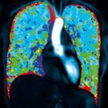
Dual-energy computed tomography (DECT) will be the main focus in cooperative project with Dr. Wolfram Stiller, physicist at the DIR. This innovative method has the potential for quantitative imaging of regional lung perfusion, and thus airway function. To accomplish this task, it is necessary to develop new models for lung perfusion and to adapt these to the conditions of DECT imaging. Subsequently, measurements in chest phantoms, animal experiments, and finally the introduction into clinical settings in patients are planned.
Research interests
- Morpho-functional MRI of human cystic fibrosis lung disease
- Functional imaging of cystic fibrosis-like lung disease in mice
- Quantitative Dual-Energy computed tomography for the modeling of lung perfusion
- Quantification of airway and parenchymal disease in obstructive lung disease
- Ex vivo porcine lung phantom