


Sektion Medizinphysik
Die erfolgreiche Strahlentherapie zeichnet sich durch eine optimale Tumorkontrolle bei geringen Nebenwirkungen aus. Dies gelingt, in dem man den Tumor möglichst präzise bestrahlt und gleichzeitig Normalgewebe und Risikoorgane optimal schont. Unter diesem Aspekt stellt die Partikeltherapie mit Schwerionen und Protonen, wie sie in Heidelberg seit 2009 am HIT angeboten wird, für viele Indikationen eine vielversprechende Behandlungsmodalität dar.
Im Zentrum unserer Forschung steht die Individualisierung der Partikeltherapie (PT) aus physikalischer Perspektive. In innovativen, interdisziplinären Querschnittsprojekten entwickeln wir aktuelle klinische Behandlungskonzepte weiter, mit dem Ziel, die vorteilhaften physikalischen Strahleigenschaften der PT in ein optimales Behandlungsergebnis für unsere Patienten zu überführen.
Aktuell bearbeiten wir zwei Schwerpunktthemen:
- Untersuchung von Dosiseffekte im zentralen Nervensystem, um daraus schonendere Bestrahlungskonzepte abzuleiten und klinisch zu etablieren
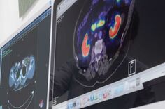
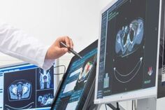
- Entwicklung und Implementierung klinischer Abläufe zur MRT-basierten bildgeführten Partikeltherapie, die in Zukunft eine engmaschige und präzise Beobachtung des Patienten unter Therapie ermöglichen.
Nähere Informationen finden sich bei den jeweiligen Mitarbeitern.
Ausbildung:
Abschlussarbeiten im Bereich Medizinphysik werden fortlaufend angeboten, bei Interesse bitte per E-Mail Kontakt aufnehmen.
LEITUNG
PD Dr. rer. nat. Julia Bauer
(kommissarisch)
WISSENSCHAFTLICHE MITARBEITER

Dr. med. Dr. rer. nat. Anika Simon
Schwerpunkt: Experimentelle Untersuchung von Strahlenwirkung im zentralen Nervensystem.
PROMOVIERENDE
M. Sc. Marc Boucsein (Dr. rer. nat.), Fakultät für Physik Heidelberg:
„Modelling of the regeneration of the central nervous system as a dynamical self-organized system“
Juliane Weber (Dr. med.), Medizinische Fakultät Heidelberg:
„Zeitliche Entwicklung klinischer Strahlenläsionen“
M. Sc. Habiba Sallem (Dr. sc. hum.), Medizinische Fakultät Heidelberg:
„Investigation of late-occurring dose effects in human brain tissue after proton irradiation“
„Radiation therapy is a pivotal approach in cancer treatment, involving the irradiation of tumor volumes with high-energy ionizing beams such as photons, protons, or heavy ions. Proton beams, owing to their unique physical characteristics, offer highly conformal and biologically effective dose delivery to tumors. While current treatment planning assumes a constant effectiveness for protons, recent findings indicate potential variability. Patients undergoing treatment for low-grade glioma may subsequently develop brain lesions, challenging conventional treatment planning. My doctoral research seeks to address this challenge by predicting and minimizing the risk of brain lesion occurrence in treatment plans, aiming for less toxicity without compromising tumor control. Additionally, my research delves into the possibilities offered by advanced MR imaging to identify early tissue changes leading to brain lesions. This innovative approach could pave the way for more targeted medication and the prevention of severe lesions. My research contributes valuable insights to cancer care and propels advancements in proton therapy through the initiation of a prospective clinical trial.“
M. Sc. Rita Pestana (Dr. sc. hum.) Medizinische Fakultät Heidelberg:
„Implementation of an MR-guided particle therapy workflow at HIT“
„Protons and heavy ions are more sensitive to changes that occur on their path compared against conventional radiotherapy with photons, making it essential to assess the patient’s anatomy along the fractions. This assessment can be done with the so-called image guided therapy: prior to some of the treatment fractions, the patient acquires an examination, usually a CT, that is analysed to assess changes in its anatomy. MRI is also a potential candidate for image-guided therapy, as it offers superior soft tissue contrast, allowing a better visualisation of the tumour. However, MRI does not provide the information required to make dose calculations (stopping power information) and, eventually, re-plan the treatment. Therefore, an additional step, the generation of a deformed CT, is required. I’m working on the clinical implementation and automation of the whole MRI-guided workflow: from the deformed CT generation to the automated re-planning, including the quantification of the uncertainties involved in each step. I’m also developing and testing tools to guide the clinicians on the identification of the anatomical changes and their impact on the prescribed treatment plan.“
Julia Hendrik (Dr. med.), Medizinische Fakultät Heidelberg:
„Latente Strahlennekrosen – Proteinregulation während der Entstehung“
MASTERSTUDIERENDE
Beatrice Geiger (M.sc.), Physikstudium an der Universität Heidelberg:
„KI-gestützte Qualitätssicherung radiotherapeutischer Bestrahlungsanlagen“
Karolin Milewski (M.sc.), Physikstudium an der Universität Heidelberg:
„Dosimetric measurements for online MR-imaging during ion beam radiotherapy“
„For very precise tumour irradiation and thereby only little complications in healthy tissue and a better tumour control, it is essential to acquire images of the patient’s anatomical structure during the treatment delivery to detect changes and adapt to them on the go. Since radiotherapy with ions already is very precise and magnetic resonance imaging (MRI) provides us with a method of radiation-free image acquisition with supreme contrast, a combination of both is a promising solution to the problem. Since the strong magnetic fields of MR devices deflect beams of charges particles on their way, more investigations on the effects of the MR device on the ion beams need to be done. I’m therefore dealing with the assurance of a reliable dose application with a charged particle beam inside an MR scanner. I’m performing dosimetric measurements in water phantoms inside an MR scanner and quantify the effects of the MRI on the dose and adapt the current standard clinical workflow for dose assurance to account for the properties of the MR scanner, using our mobile MR device installed at the experimental beamline of the Heidelberg Ion-Beam Therapy Center (HIT). I then compare the measured dose to computational dose calculations to ensure dose consistency. With this project the combination of real-time MRI and ion beam radiotherapy comes on step close to clinical reality.“
Lotta Solle (M.sc.), Medizinphysikstudium an der Hochschule für Wissenschaft und Technik Saarbrücken:
„Untersuchung der Lagerungsgenauigkeit in einer drehbaren Patientenkapsel für die bildgeführte Partikeltherapie“
“Ich verfasse meine Masterarbeit zu dem Schwerpunkt MRT-basierte Plananpassung für eine MR-gesteuerte Gantry-lose Partikeltherapie. Anstatt den Patienten mithilfe einer Gantry aus verschiedenen Richtungen zu bestrahlen, soll der Patient selbst gedreht werden. Zur Drehung des Patienten vor einem horizontalen Strahl wurde ein Patientenrotationssystem, auch Patientenkapsel genannt, entwickelt. Genauer widme ich der Fragestellung, wie präzise die Lagerung eines Patienten in dieser Patientenkapsel möglich ist und wie reproduzierbar die Positionierung für eine Ionenstrahltherapie in einer solchen Kapsel ist. Im Rahmen einer Probandenstudie wurden bereits MRT-Aufnahmen in verschiedenen Lagerungswinkeln (0°, 70°, 90° und 110°) in mehreren Durchläufen erstellt. Im Rahmen meiner Masterarbeit vergleiche ich nun die verschiedenen MRT-Aufnahmen hinsichtlich der Bewegung des Probanden relativ zur Patientenkapsel sowie der Bewegung der Organe relativ zu einer festen Knochenstruktur wie der Wirbelsäule.”
EHEMALIGE MITARBEITER
Dr. rer. nat. Robin Koch:
Dissertation 11/2023, Fakultät für Biowissenschaften Heidelberg, “Statistical analysis and modelling of time-resolved In Vitro Clonogenic Assays“
Prof. Dr. rer. nat. Markus Alber:
Sektionsleitung (bis 03/2023)
Dr. rer. nat. Emanuel Bahn:
wissenschaftlicher Mitarbeiter (bis 03/2023)
M. Sc. Paul Meder:
wissenschaftlicher Mitarbeiter (bis 01/2023)
M. Sc. Teresa Rodriguez Gonzales, Promotion in Physik an der Universität Sevilla, Forschungsaufenthalt (09-12/2021):
„Towards PET range verification in proton therapy: new cross sections for improved accuracy“
M. Sc. Judith Besuglow:
Physikstudium an der Universität Heidelberg, Masterarbeit (01/18-04/19): „Design präklinischer in vivo Präzisions-Bestrahlung“
Christoph Harmel:
Biotechnologie-Student an der Universität Heidelberg, Praktikum (07/18-10/18): „Zeitaufgelöster In Vitro Clonogenic Assay“
PUBLIKATIONEN
FORSCHUNGSFÖRDERUNG
Unsere Forschung wird finanziell unterstützt durch:
DFG
BMBF
Universität Heidelberg (Olympia-Morata-Programm)
Alois-Hirdt Erben und Wieland Stiftung Heidelberg