


Forschungsgruppe Neue Detektionstechniken für Ionenstrahlen
Klinische Studie InViMo
In-vivo monitoring of carbon ion radiotherapy delivery
Die Strahlentherapie mit Kohlenstoffionen, die an dem Heidelberger Ionentherapie Zentrum durchgeführt wird, ist ein hochpräzises Verfahren zur Behandlung von bestimmten Krebsarten, die mit der Standardtherapie schwer oder gar nicht behandelbar sind (z.B. Tumore der Schädelbasis). Gegenüber der Strahlentherapie mit Photonen sind die Kohlenstoffionenbestrahlungen empfindlicher gegenüber Änderungen im Patienten, wie z.B. eine mögliche Verkleinerung des Tumorvolumens im Verlauf der Therapie. Daher ist die Überwachung des Ionenstrahls im Patienten während der Behandlung von großem Interesse.
Warum die InViMo Studie?
Der therapeutische Kohlenstoffionenstrahl bremst im Patienten ab und wirkt hauptsächlich im Tumor. Während der Therapie, wie bei jeder Art von Strahlentherapie, tritt aus dem Patienten Sekundärstrahlung aus. Die Idee der InViMo Studie ist, sich diesen Nebeneffekt zu Nutze zu machen, um den Kohlenstoffionenstrahl im Patienten zu überwachen.
In der InViMo Studie wird untersucht, in wie weit die Sekundärstrahlung bei der Strahlentherapie mit Kohlenstoffionen Informationen beinhaltet, die Rückschlüsse auf kleine Änderungen des Tumorvolumens bereits während des Therapieverlaufs erlauben.
Das Verfahren hat sich in Voruntersuchungen bereits als vielversprechend erwiesen. In dieser Studie wird das Potential dieses neuen Verfahrens an Patienten untersucht.
Mit der Teilnahme an der Studie sind keine zusätzlichen Risiken oder Nebenwirkungen verbunden. Es erfolgt weder eine zusätzliche Bildgebung, noch besteht eine zusätzliche Strahlungsexposition des Patienten. Ebenfalls gibt es keinen Einfluss auf die Strahlentherapie.
Wie läuft die klinische Studie ab?
Im Rahmen der Studie finden während der Strahlenbehandlung kontaktlose Messungen der Sekundärstrahlung, die aus dem Patientenkörper austritt, statt.
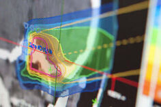
Anhand der gesammelten Daten werten wir potentielle Änderungen des Strahlungsfeldes im Patienten aus, die z.B. auf Änderungen des Tumorvolumens hinweisen könnten. Die Resultate der neuen Methode werden mit denen der CT Bildgebung verglichen, die standardmäßig als Kontrolle während des Therapieverlaufes erfolgt. Es entsteht somit keine zusätzliche Strahlenbelastung des Patienten.
Von dieser Studie haben die teilnehmende Patienten selbst keinen direkten persönlichen Nutzen. Die Resultate helfen uns die Therapie eventuell zu verbessern und somit zukünftigen Patienten besser zu helfen.
Was passiert während der Bestrahlungssitzung?
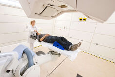
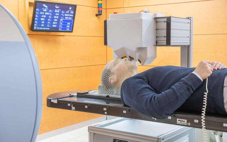
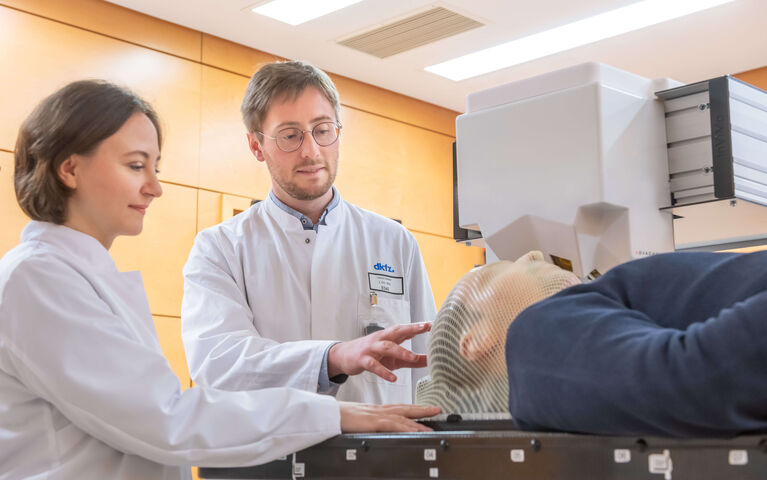
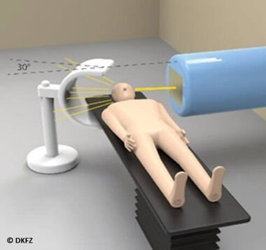
Wie bei den Bestrahlungssitzungen üblich ist, wird zuerst der Patient auf der Patientenliege positioniert. Anschließend wird ein Messgerät neben der Liege platziert, das die Sekundärstrahlung, die aus dem aus dem Patienten austritt, aufzeichnen kann (siehe Abbildung 1). Die Messung der Sekundärstrahlung erfolgt kontaktlos und ohne den Einsatz zusätzlicher Strahlung.
Mit der Teilnahme an der Studie beläuft sich der zeitliche Mehraufwand auf weniger als 5 Minuten pro Therapiesitzung. Die Zeit wird benötigt, um das Messsystem vor der Bestrahlung neben dem Patiententisch zu platzieren und nach dem Abschluss der Bestrahlung wieder zu entfernen. Innerhalb der Studie sind Messungen bei bis zu 10 Bestrahlungsterminen vorgesehen. Somit beträgt der zeitliche Gesamtaufwand für den Patienten insgesamt weniger als eine Stunde während der gesamten Studie. Es sind keine zusätzlichen Termine oder Untersuchungen geplant.
Ein weltweit einzigartiges Messgerät
Das Messgerät ist eine weltweit einmalige Art von Kamera. Es ist ein Paradebeispiel der Übertragung von Wissen aus der Grundlagenforschung hin zur Verbesserung der Behandlung der Krebspatienten. Das Messgerät basiert auf jahrzehntelanger Entwicklung, die am Forschungszentrum CERN in Genf in der Schweiz ihren Anfang nahm. Ursprünglich wurde diese Art von Strahlungsdetektoren, sogenannte Pixeldetektoren die aus Halbleitern bestehen, in den größten Experimenten der Welt eingesetzt um bis dahin unbekannte Teilchen der Materie zu finden. Ein Beispiel ist das berühmte Higgs Boson. Die Medipix Kollaboration am CERN hat die Technologie für ein breites Spektrum von Forschungsprojekten zugänglich gemacht. Der Kern des Messgerätes wurde von der Firma ADVACAM s.r.o. aus Prag, Tschechien, geliefert. Insgesamt beruht das Messverfahren auf mehr als 10-jähriger Forschung und Entwicklung durch unser Team, die in namhaften wissenschaftlichen Zeitschriften publiziert wurde.
Die Studie wird gefördert durch:
Program Proof-of-Concept Clinical Studies

Die Partner der klinischen Studie sind:

Der Kern des Messgerätes wurde geliefert von der Firma:
Medizinische Leitung der Studie:
Universtiätsklinikum Heidelberg
Klinik für Radioonkologie und Strahlentherapie
Dr. Semi Harrabi, Oberarzt
Prof. Dr. Dr Jürgen Debus, Ärztlicher Direktor
Physikalische Leitung und Entwicklung des Messgerätes:
Deutsches Krebsforschungszentrum, Heidelberg
Deutsches Krebsforschungszentrum, Heidelberg
Abteilung Medizinische Physik in der Strahlentherapie
Dr. Maria Martisikova, Gruppenleiterin
Prof. Dr. Oliver Jäkel, Abteilungsleiter
Wissenschaftliche Veröffentlichungen zum Thema:
1. L. Ghesquière-Diérickx, R. Félix-Bautista, A. Schlechter, L. Kelleter, M. Reimold, G. Echner, P. Soukup, O. Jäkel, T. Gehrke and M. Martišíková. Detecting perturbations of a radiation field inside a head-sized phantom exposed to therapeutic carbon-ion beams through charged-fragment tracking. Medical Physics 29 (2022) 1776-1792
2. L. Ghesquière-Diérickx, A. Schlechter, R. Félix-Bautista, T. Gehrke, G. Echner, L. Kelleter and M. Martišíková. Investigation of Suitable Detection Angles for Carbon-Ion Radiotherapy Monitoring in Depth by Means of Secondary-Ion Tracking. Frontiers in Oncology 11 (2021) 780221
English
InViMo clinical study:
In-vivo monitoring of carbon-ion radiotherapy delivery
Radiotherapy with carbon ions, as it is performed at the Heidelberg Ion Beam Therapy Center, is a highly precise treatment modality for tumors that can not easily be treated with conventional X-ray radiotherapy (e.g. tumors at the base of skull). Compared with X-ray radiotherapy, carbon-ion beams are more sensitive to changes in the patient anatomy such as a shrinkage of the tumor volume over the course of the treatment. Therefore, ion-beam monitoring in the patient (in-vivo) during treatment is of great interest.
Why do we do the InViMo study?
The therapeutic carbon-ion beam gradually slows down in the patient and is most effective in the tumor. Just as in any other form of radiotherapy, there is secondary radiation emerging from the patient. The principle of InViMo is to use this side effect to monitor the carbon-ion beam in the patient.
In the InViMo study it is investigated if the secondary radiation carries information that can be used to draw conclusions about small tumor volume changes during the treatment.
The method showed promising results in previous studies. In this study, the potential of the method is investigated in patients.
The participation in this study comes without additional risks or side effects to the patient. There is no additional imaging nor radiation exposure. Moreover, there is no modification of the regular treatment course.
What is the clinical workflow?
In the course of the study, contactless measurements of the secondary radiation, which emerges from the patient, is performed. Based on this data, observed differences in the secondary radiation field are traced back to potential changes of the tumor volume. The results of this new method are compared to control CT images, which are acquired as part of the regular treatment course. Therefore, there is no additional radiation exposure of the patient.
Patients do not have an immediate personal gain from the participation in the InViMo study. Instead, the results will help us to potentially improve the treatment of future patients.
What happens during a treatment fraction?
As usual in radiotherapy, the first action of a treatment fraction is to position the patient on the patient table. Afterwards, a detection system that measures the secondary radiation emerging from the patient, is placed next to the patient table (see figure 1). The measurement of the secondary radiation happens contactless and without any additional radiation exposure.
The extra time that comes with the participation in the InViMo study is less than 5 minutes per treatment fraction. The time is needed to place the detection system next to the patient table before the treatment and to remove it afterwards. Up to a maximum of 10 treatment fractions will be monitored. Therefore, the total extra time for a participating patient is less than one hour over the course of the entire study. No additional appointments or examinations are planned.
A worldwide unique detection system
The detection system is best compared to a worldwide unique type of camera. It is a prime example of knowledge transfer from fundamental research to the improvement of cancer therapy. Decades of research and development were required to build the detection system, which began at the research center CERN . Initially, the semiconductor pixel detectors used here were developed to find yet unknown elementary particles with the help of the largest experiments ever built. A famous example of such an elementary particle is the Higgs boson. The Medipix collaboration at CERN made this technology available to a large variety of research projects. The principal components of the detection system were supplied by the company ADVACAM s.r.o. (Prague, Czech Republic). More than 10 years of research and development in our team at DKFZ preceded the completion of the detection system, of which the milestones were published in renewed scientific journals .
Contact
Medical Physics in Radiation Oncology E040
German Cancer Research Center
Im Neuenheimer Feld 280
69120 Heidelberg
Tel: (+49) 06221 42 2441
E-Mail: m.martisikova(at)dkfz.de