![[Translate to English:]](/fileadmin/_processed_/8/2/csm_20131204_Beratung_035_a396c6c6e5.jpg "[Translate to English:]")
![[Translate to English:]](/fileadmin/_processed_/a/0/csm_20170627_PflegeOrtho_001_fb912471fa.jpg "[Translate to English:]")
![[Translate to English:]](/fileadmin/_processed_/f/c/csm_20170215_LaborOMZ_155_c0169c0898.jpg "[Translate to English:]")
![[Translate to English:]](/fileadmin/_processed_/2/c/csm_20180523_Labor_139_6ebb9a0a1b.jpg "[Translate to English:]")

Detection of numerical chromosomal abnormalities
(FISH interphase )
By hybridizing specific DNA probes – ideally from the centromere region of chromosomes – specific chromosome copy-number alterations can be detected directly at the cell nucleus.
Detection of structural chromosomal aberrations
Apart from commercial probe sets, our laboratory makes use of a broad range of different DNA probes to visualize whole chromosomes (chromosome-specific coloring-probes) and chromosomal subregions (probes for the subtelomeric regions, single genes and specific bands). Due to our extensive experience in the use of multicolor FISH protocols, we are able to hybridize up to 7 different probes at the same time.
Microdeletion syndromes
Williams-Beuren syndrome: FISH analysis with a probe set for chromosome 7q11.2 (ELN/LIMK1/D7S613, red) and a reference probe for chromosome 7q31 (D7S486/D7S522, green) showing a microdeletion 7q11.2 in chromosome 7.
Table: Microdeletion syndromes
Analysis of subtelomeres
In 5-10% of all patients with psychomotor retardation and dysmorphia or organ malformations of unknown origin, we suspect the cause to be alterations at the chromosome ends which are not visible under a light microscope. In subtelomere analysis, BAC and PAC clones are used as probes to visualize all chromosome ends (except the short arms of acrocentric chromosomes) for FISH analysis. Due to the extensive testing required, karyotyping (resolution: approximately 500 bands) should be performed beforehand and the test indication should be made in a genetic counseling session.
Prenatal rapid test
Prenatal rapid test: Hybridization of probe set (1) for chromosomes 13 (LSI 13, green) and 21 (LSI 21, red) and probe set (2) for chromosomes X (DXZ1, green), Y (DYZ3, red), and 18 (D18Z1, blue) showing an inconspicuous female karyotype in the amniotic fluid nuclei for the above probes.
FISH interphase analysis on amniotic fluid cell nuclei allows to quickly identify the most common numerical chromosomal abnormalities in humans (trisomies of chromosomes 21, 18, and 13, and numerical abnormalities of sex chromosomes e.g. monosomy X and Klinefelter syndrome). The results are usually returned the next day. However, a prenatal rapid test cannot replace formal karyotyping as it is not able to detect all chromosomal structural abnormalities. Particulary in case of chromosomal mosaics, the diagnostic value of the rapid test is limited. Prenatal rapid tests are not covered by health insurance and must be billed as individual health service (IGeL).
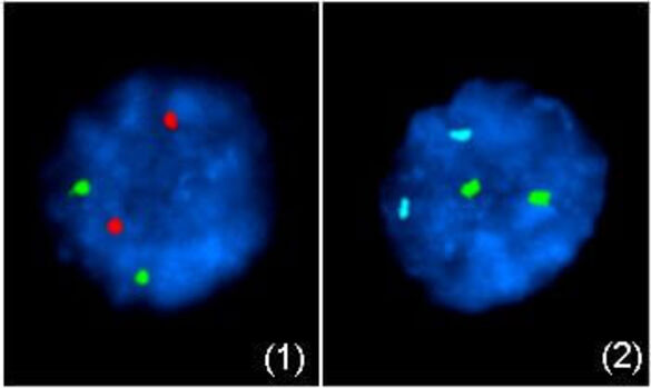
Tumor cytogenetics (FISH interphase)
Numerous malignant hematological diseases exhibit numerical and/or structural chromomosomal aberrations that are diagnostically, prognostically and therapeutically relevant. For cytogenetic studies, the preparation of metaphase chromosomes is essential, but the spontaneous division activity of leukemic cells in vitro is not always sufficient for this purpose. By hybridizing specific tumor-relevant DNA probes, the diagnosis of numerical (image) and structural chromosomal aberrations (image) is also possible directly in the cell nucleus (FISH interphase).
Table: Hematological diseases
For more information please contact Prof. Dr. A. Jauch (Tel.: +49 6221 56-5407 / -5063).
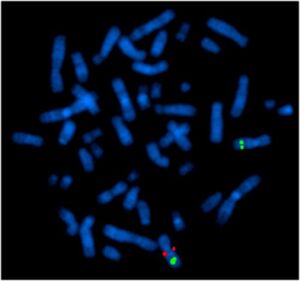
Multiplex fluorescence in situ hybridization (M-FISH)
The M-FISH technique allows the simultaneous visualization of all human chromosomes in 24 different colors. The method is based on a combinatorial labeling strategy of chromosome-specific paint probes. Using 7 different fluorochromes, each chromosome can be identified by the defined color or color combination. Indications for this relatively complex technique are the identification of complex aberrant karyotypes (image) or the chromosomal identification of marker chromosomes (exception: heterochromatic markers and low grade mosaics).
Please contact Prof. Dr. A. Jauch (Tel.: +49 6221 56-5407 / -5063) before submitting a test request.