


CLINICAL TRIAL SERVICES
We are an innovation-oriented partner in the field of hemato-immuno-oncological diagnostics and accompanying immune-monitoring in the context of preclinical and clinical trials.
We are happy to assist you with reliable flow cytometry and routine molecular diagnostics together with our innovations from the Clinical Immunology Laboratory. Modern approaches and many years of cooperation with scientific institutions and pharmaceutical companies underline our expertise in the development of unique and validated methods for potential immunotherapies. From target identification to clinical evaluation - our clinical immunology laboratory provides quantitative methods for immunotherapy monitoring using in vitro models. This also includes the use of transcriptional high-throughput sequencing approaches to assess the effect of immunotherapy at the molecular level.
Head of the Institute

PROJECT MANAGEMENT
Specialization: Immunology, Clinical Pharmacology and Molecular Biology
Specialization: Flow Cytometry, Quality Management
Specialization: Flow Cytometry, Biobanking, Biostatistics, Project management, Project Funding, Regulatory Requirements
Specialization: Molecular Genetics
Specialization: Flow Cytometry
SPECTRUM OF METHODS
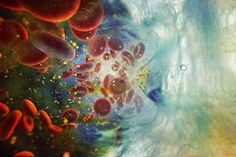
Flow Cytometry
Flow Cytometry allows us to characterize a large number of single cells in a short amount of time regarding their surface and intracellular antigens. Using this high throughput method allows us to present complex results from various sample sources (including blood, bone marrow, cerebrospinal fluid and solid tissues after preparation).
MULTIPARAMETER FLOW CYTOMETRY
The bandwidth of hematological malignancies requires an analysis method to characterize the complex biomarker combinations needed for a diagnosis. We use modern 8-10 color flow cytometers which are capable of performing these multiparametric analyses.
NUMBER OF SITES QUANTIFICATION
By using a special combination of antibody and analysis we are able to give an exact number of a particular surface marker per single cell. This knowledge can serve as a prognostic factor in drug evaluation.
IMMUNOPROFILING
By describing the composition of the patient’s immune system, we can support and learn more about immune modulatory therapies (IMID) in hematological malignancies.
TARGETING
By characterizing the cells’ surface markers we can detect targets for new chemotherapies, new drug conjugated antibodies and CAR-T-cell therapies on cell samples of relapsed patients and are able to give a fast feedback to the clinicians concerning the possibility for the use of these new promising therapy options.
Together with our Clinical Immunology Lab we develop possible new targets.
NEXT GENERATION FLOW
The detection of minimal residual disease requires cutting edge technology to detect very small numbers of residual cells of the primary malignancy. After analyzing the cells in our modern flow cytometers, we use highly innovative semi-automated software tools to further characterize the samples. This method allows a prognostic assessment early on.
Clinical Immunology
ELISPOT
The enzyme-linked immune absorbent spot is a well-established method used to quantitatively measure the secretion of specific cytokines from immune cells. We utilize the method to accurately assess the effect of drugs (or even gene knock down methods) on the function of immune cells using Tumor-antigen-expressing cells as surrogate targets.
RNA Sequencing
Using next generation sequencing approaches, we examine the quantity of tens of thousands of genes simultaneously. Such an approach provides very valuable and accurate information on the effect of the experimental probes on every cellular compartment.
CRISPR Cas9 Knock Down
Using the recent advances in gene knock down methods, we use the Cas9-based knock down approach in order to investigate the function of specific genes-of-interest and to evaluate possible immune check points in tumor immunology.
Luciferase Reporter Assay
Luciferase reporter assay is a widely used approach to explore the role of transcriptional factors in regulating specific protein expression. In tumor immunology, the Luciferase assay provides important information about the role of transcriptional factors in regulating the cellular fate.
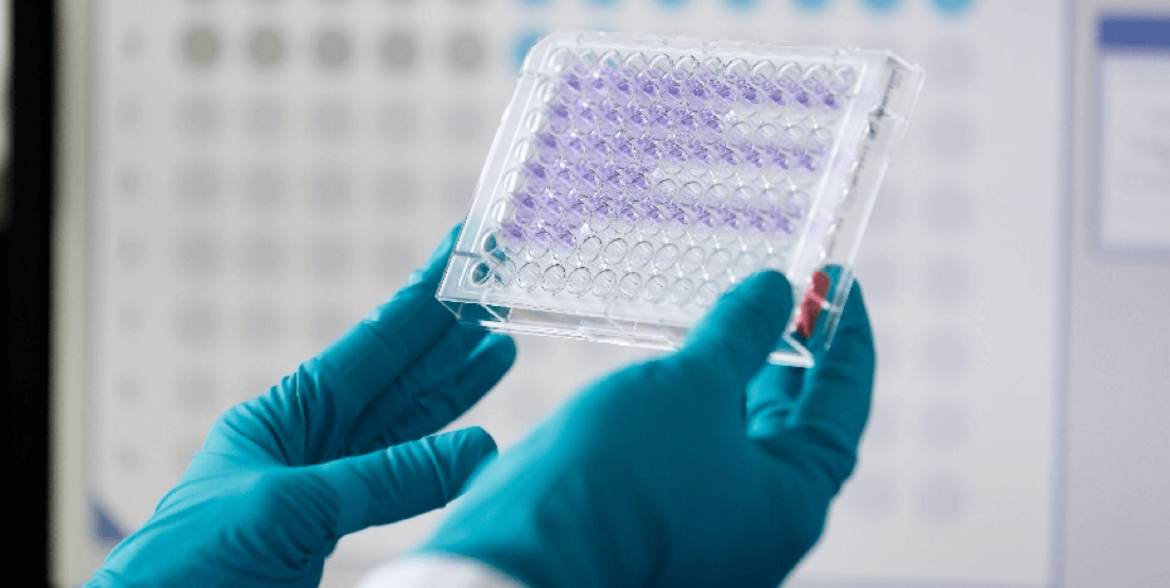
ELISA
The enzyme-linked immunosorbent assay is a standard laboratory approach to quantify the secretion of cytokines from immune cells with very high sensitivity (as low as a few nanograms per milliliter)
Molecular Genetics
Based on the mutation(s) of interest we are able to select the appropriate method (see below) for analysis and to establish the analysis including suitable controls.
PCR (polymerase chain reaction)
PCR is our basic method used to amplify regions of interest which can be analyzed further using various approaches (see below). Specificity of the amplified products is based on the selection of specific primers. During PCR the region of interest is amplified exponentially which allows further analysis as well as the detection of rare events.
Restriction
Restriction enzymes can be used to detect mutations if the mutation leads to the loss of a specific restriction site. Based on the position of the primers used for PCR the resulting band pattern on an agarose gel or peaks on a capillary sequencer differs between wildtype- and mutant-DNA.
Multi-plex PCR
Since genomic rearrangements can occur within different breakpoint regions of genes, multi-plex PCR uses various primers covering different breakpoint regions in a single PCR reaction. This enables us to detect different genomic rearrangements very efficiently. This method is used to detect genomic rearrangements not necessarily leading to an enhanced expression and short intronic sequences.
Long Range PCR
This method allows the amplification of genomic rearrangements leading to PCR products up to 20 kb. This method is used to detect genomic rearrangements not necessarily leading to an enhanced expression and long intronic sequences.
RT-PCR (reverse transcription PCR)
RT-PCR is used to detect genomic rearrangements based on RNA. Since intronic sequences which can be very long are only present in genomic DNA but absent in mRNA, using RNA, which has to be reverse transcribed into cDNA, as template allows to amplify genomic rearrangements more efficiently. This method is used for the detection of genomic rearrangements with large intronic sequences where the genomic rearrangement leads to an enhanced expression of the fusion gene.
Real-time PCR
Real-Time PCR is used to quantify specific transcripts either to measure expression levels of genes of interest or to calculate the amount of specific transcripts compared to a reference gene. Target detection is based on fluorescence signals which are measured at the end of every single PCR cycle. Dependent on the amount of target present in the real-time PCR reaction the fluorescence signal crosses the determined threshold at different PCR cycles. The PCR cycle at which the threshold is crossed is used for quantification.
Allele-specific PCR
Allele-specific PCR is a method to detect point mutations. For this, a primer specific for the wildtype allele and a primer specific for the mutant allele as well as a primer beneath the mutation site which binds to both, the wildtype as well as the mutant allele is used. In order to detect the different alleles two PCR reactions, one using the wildtype-specific and one using the mutant-specific primer, are performed. By gel electrophoresis (see below) the presence of the different alleles in the sample can be analyzed.
ARMS-PCR (Amplification Refractory Mutation System-PCR)
ARMS-PCR is a kind of allele-specific PCR allowing the detection of different alleles in one sample. Using an ARMS-PCR approach the wildtype and mutant allele can be detected in a single PCR reaction. In addition to the allele-specific primers also two common primers are used leading to a PCR product which is present independent of the presence/absence of the mutant allele. For ARMS-PCR the position of the common primers has to be selected in a way that the PCR-products resulting from the wildtype or the mutant allele are of different size. By gel electrophoresis (see below) the presence of the different alleles in the sample can be analyzed based on the different fragment sizes of wildtype and mutant PCR-products.
Digital droplet PCR
Digital droplet PCR is a method for detection and quantification of rare events by fluorescence. Due to its very high sensitivity compared to Sanger Sequencing digital droplet PCR can be used to monitor minimal residual disease as well as to determine allelic frequencies of mutations which might have a prognostic impact.
Fragment sizing
Fragment sizing is used to detect insertions or deletions within genomic regions. For this at least one fluorescence-tagged primer is used for PCR. The PCR products are then analyzed on a capillary sequencer in order to determine the precise size(s) of the resulting PCR-product(s). Due to insertions or deletions the sizes differ between wildtype- and mutation-DNA. Using this method, it is also possible to determine allelic ratios of wildtype and mutant.
Sanger Sequencing
Sanger Sequencing is used to determine the nucleotide sequence of a region of interest. This method is suitable if there might be mutations at different nucleotides within a defined region as well as to determine the precise mutation if there various mutations possible at a single nucleotide position.
NGS (Next Generation Sequencing)
Next generation sequencing is employed to analyze the nucleotide sequence of various genes or parts of various genes of different patients in a single experiment. By this, a huge amount of sequencing data is generated in order to identify mutations within different genes which might contribute to disease development or allows a prognostic assessment.
OUR EXPERTISE
The following preclinical and clinical trials were and are accompanied by our laboratory with minimal residual disease assessment and advanced immune profiling portfolio:
- GMMG HD6, multiple myeloma phase III clinical trial Studie, multicentric
- GMMG HD7, multiple myeloma phase III clinical trial Studie, multicentric
- GMMG CONCEPT, multiple myeloma phase III clinical trial, multicentric
- Sanofi CD38-Number-of-sites-Analysis
- Alexion PNH study
- Multiple Myeloma Research Foundation (MMRF) Research Cooperative „Immune Regulation and Dysfunction within the Bone Marrow in Multiple Myeloma“
- SAL AML TEAM trial
OUR PUBLICATIONS
K. Kriegsmann, M.-A. Baertsch, M.H.S. Awwad, M. Merz, D. Hose, A. Seckinger, A. Jauch, N. Becker, A. Benner, M.S. Raab, J. Hillengass, U. Bertsch, J. Dürig, H.J. Salwender, M. Hänel, R. Fenk, M. Munder, K. Weisel, C. Müller-Tidow, H. Goldschmidt, M. Hundemer, Cereblon-binding proteins expression levels correlate with hyperdiploidy in newly diagnosed multiple myeloma patients, Blood Cancer J. 9 (2019)
K. Kriegsmann, M. Kriegsmann, M. Cremer, M. Schmitt, P. Dreger, H. Goldschmidt, C. Müller-Tidow, M. Hundemer, Cell-based immunotherapy approaches for multiple myeloma, Br. J. Cancer 120 (2019)
M.H.S. Awwad, K. Kriegsmann, J. Plaumann, M. Benn, J. Hillengass, M.S. Raab, U. Bertsch, M. Munder, K. Weisel, H.J. Salwender, M. Hänel, R. Fenk, J. Dürig, C. Müller-Tidow, H. Goldschmidt, M. Hundemer, The prognostic and predictive value of IKZF1 and IKZF3 expression in T-cells in patients with multiple myeloma, Oncoimmunology 7 (2018)
Kriegsmann K, Löffler H, Eckstein V, Schulz R, Kräker S, Braun U, Luft T, Hegenbart U, Schönland S, Dreger P, Krämer A, Ho AD, Müller-Tidow C, Hundemer M, CD7 is expressed on a subset of normal CD34-positive myeloid precursors. Eur J Haematol (2018)
Kriegsmann K, Dittrich T, Neuber B, Awwad MHS, Hegenbart U, Goldschmidt H, Hillengass J, Hose D, Seckinger A, Müller-Tidow C, Ho AD, Schönland S, Hundemer M, Quantification of number of CD38 sites on bone marrow plasma cells in patients with light chain amyloidosis and smoldering multiple myeloma. Cytometry B Clin Cytom. (2018)
J. Plaumann, M. Engelhardt, M.H.S. Awwad, H. Echchannaoui, E. Amman, M.S. Raab, J. Hillengass, N. Halama, B. Neuber, C. Müller-Tidow, H. Goldschmidt, M. Hundemer, IL-10 inducible CD8+ regulatory T-cells are enriched in patients with multiple myeloma and impact the generation of antigen-specific T-cells, Cancer Immunol. Immunother. 67 (2018)
Lisenko K, Schönland S, Hegenbart U, Wallenwein K, Braun U, Mai EK, Hillengass J, Goldschmidt H, Jauch A, Ho AD, Raab M, Hundemer M, Potential therapeutic targets in plasma cell disorders: A flow cytometry study. Cytometry B Clin Cytom. (2017)
Neuber, J. Dai, W.A. Waraich, M.H.S. Awwad, M. Engelhardt, M. Schmitt, S. Medenhoff, M. Witzens-Harig, A.D. Ho, H. Goldschmidt, M. Hundemer, Lenalidomide overcomes the immunosuppression of regulatory CD8+CD28- T-cells, Oncotarget 8 (2017)
A. Seckinger, J.A. Delgado, S. Moser, L. Moreno, B. Neuber, A. Grab, S. Lipp, J. Merino, F. Prosper, M. Emde, C. Delon, M. Latzko, R. Gianotti, R. Lüoend, R. Murr, R.J. Hosse, L.J. Harnisch, M. Bacac, T. Fauti, C. Klein, A. Zabaleta, J. Hillengass, E.A. Cavalcanti-Adam, A.D. Ho, M. Hundemer, J.F. San Miguel, K. Strein, P. Umaña, D. Hose, B. Paiva, M.D. Vu, Target Expression, Generation, Preclinical Activity, and Pharmacokinetics of the BCMA-T Cell Bispecific Antibody EM801 for Multiple Myeloma Treatment, Cancer Cell 31 (2017)
Lisenko K, Schönland SO, Jauch A, Andrulis M, Röcken C, Ho AD, Goldschmidt H, Hegenbart U, Hundemer M, Flow cytometry-based characterization of underlying clonal B and plasma cells in patients with light chain amyloidosis.Cancer Med. (2016)
I. Krämer, M. Engelhardt, S. Fichtner, B. Neuber, S. Medenhoff, U. Bertsch, J. Hillengass, M.-S. Raab, D. Hose, A.D. Ho, H. Goldschmidt, M. Hundemer, Lenalidomide enhances myeloma-specific T-cell responses in vivo and in vitro, Oncoimmunology 5 (2016)
S. Fichtner, D. Hose, M. Engelhardt, T. Meißner, B. Neuber, F. Krasniqi, M. Raab, S. Schönland, A.D. Ho, H. Goldschmidt, M. Hundemer, Association of antigen-specific T-cell responses with antigen expression and immunoparalysis in multiple myeloma, Clin. Cancer Res. 21 (2015)
M. Munder, M. Engelhardt, D. Knies, S. Medenhoff, G. Wabnitz, C. Luckner-Minden, N. Feldmeyer, R.-H. Voss, P. Kropf, I. Müller, R. Conradi, Y. Samstag, M. Theobald, A.D. Ho, H. Goldschmidt, M. Hundemer, Cytotoxicity of tumor antigen specific human T cells is unimpaired by arginine depletion, PLoS ONE 8 (2013)
COLLABORATIONS
- University Medicine Mainz (AG Echchannaoui)
- DKFZ/Division of Applied Bioinformatics (AG Brors)
- DKFZ/Biostatistics (Axel Brenner)
- EMBL (Genomics and Proteomics Core Facilities)
- GMMG Study Group
- Cytognos – Flow Cytometry Solutions (Salamanca, Spain)
- University of Salamanca/Flow Cytometry in Multiple Myeloma (Alberto Orfao)
- University of Navarra/ Flow Cytometry in Multiple Myeloma (Bruno Paiva)